Stickler syndrome

Osteochondritis
dissecans and Osgood Schlatter disease in a family with Stickler syndrome. Patient 1: Vertebral column lateral radiogram at age of 15 years showed platyspondyly with anterior end-plate irregularity associated with irregular surfaces of the superior and inferior apophyses.

Osteochondritis
dissecans and Osgood Schlatter disease in a family with Stickler syndrome. Patient 1: Pelvis Anteroposterior radiograph at age of 15 years showed coxa valga, relative flattening of capital femoral epiphyses, joint space narrowing and irregularity, with features of left acetabular osteochondritis associated with necrotic adjacent femoral epiphysis (arrows).

Osteochondritis
dissecans and Osgood Schlatter disease in a family with Stickler syndrome. Patient 1: Axial CT scan at age of 17 years shows a necrotic acetabular roof (arrow).

Osteochondritis
dissecans and Osgood Schlatter disease in a family with Stickler syndrome. Patient 1: Anteroposterior knee radiograph at age of 15 years shows osteochondritis dissecans of the distal femoral epiphysis with areas of evident epiphyseal necrosis (arrows).

Osteochondritis
dissecans and Osgood Schlatter disease in a family with Stickler syndrome. Patient 1: Coronal MR image of the knee at age of 17 years shows progressive and lesion from the underlying subchondral bone with apparent anterior separation of the articular surface with evidence of epiphyseal friability. Note the bony fragment had resulted in a concave crater on the femoral condyle with steeply sloping edges (arrows)

Osteochondritis
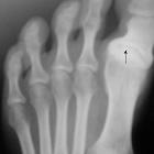
dissecans and Osgood Schlatter disease in a family with Stickler syndrome. Patient 1: Anteroposterior at age of 17 years foot radiograph showed progressive hallux varus with marked osteoarthritis of the base of the first toe.

Osteochondritis
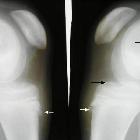
dissecans and Osgood Schlatter disease in a family with Stickler syndrome. Patient 2: Lateral radiograph of the knee with the leg internally rotated (10–20°) showed bilateral irregularity of the apophysis with separation from the tibial tuberosity.
Stickler syndrome refers to a group of disorders primarily affecting connective tissue.
Pathology
Several gene mutations have been identified dependent on specific subtypes which include:
- Stickler syndrome type I: COL2A1
- Stickler syndrome type II: COL9A1
- Stickler syndrome type III: COL11A1
- autosomal recessive Stickler syndrome: COL11A2
Clinical spectrum
Described features include:
Ophthalmologic
- congenital or early-onset cataract
- congenital vitreous anomaly, rhegmatogenous retinal detachment
- myopia greater than -3 diopters
Craniofacial
- midface hypoplasia
- depressed nasal bridge
- anteverted nares: characteristic facies typically more pronounced in childhood
- bifid uvula
- cleft hard palate
- micrognathia
- Robin sequence: micrognathia, cleft palate, glossoptosis
Audiologic
- sensorineural or conductive hearing loss
- hypermobile middle ear systems
Skeletal
- joint hypermobility
- mild spondyloepiphyseal dysplasia
- precocious osteoarthritis
Siehe auch:
und weiter:

 Assoziationen und Differentialdiagnosen zu Stickler-Syndrom:


