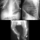
micrognathia

The term micrognathia describes a small mandible.
Epidemiology
Associations
Micrognathia is associated with a vast array of other congenital anomalies which include:
- aneuploidic syndromic
- non-aneuploidic syndromic
- Fryns syndrome
- Goldenhar syndrome: hemifacial microsomia
- hydrolethalus syndrome
- lethal multiple pterygium syndrome
- Nager syndrome
- Neu-Laxova syndrome
- Pena-Shokeir syndrome
- Pierre Robin syndrome
- Seckel syndrome
- Smith-Lemli-Opitz syndrome
- Stickler syndrome
- TAR syndrome
- Treacher Collins syndrome: mandibulofacial dysostosis
- non-syndromic non-aneuploidic
Pathology
A small mandible occurs secondary to abnormalities of the first branchial arch which in turn are caused by deficient or insufficient migration of neural crest cells and usually occur around the 4 week of gestation.
Radiographic features
Antenatal ultrasound
Due to a large portion of normal mandibular growth occurring in the 3 trimester, the condition is best diagnosed towards the latter half of pregnancy
- a true sagittal facial image would show a receding chin
- the facial profile view is most useful in evaluating the mandibular size
Micrognathia is often a subjective finding best appreciated on a midline sagittal view.
Parameters used for objective measurement include:
- jaw index: (mandibular anteroposterior diameter/biparietal diameter) x 100
- frontal nasomental angle
Ancillary sonographic features
If fetal swallowing is impaired there may be evidence of polyhydramnios.
Significance
Due to a high association rate with other anomalies, the detection of micrognathia warrants a careful search for other fetal abnormalities.
Treatment and prognosis
The overall prognosis is highly variable dependent on the presence of other associated anomalies. Even when there is isolated fetal breathing (respiratory) difficulty at the time of birth, it is a concern . In selected case a genioplasty may be an option in later life.
Complications
Severe micrognathia can potentially compromise neonatal respiration after birth.
See also
Siehe auch:
- Pätau-Syndrom
- Ehlers-Danlos syndrome
- Herzfehler
- Marfan-Syndrom
- Turner-Syndrom
- Polyhydramnion
- Trisomie 18
- mandibuläre Retrognathie
- Embryopathia rubeolosa
- Franceschetti-Zwahlen-Syndrom
- Pena-Shokeir-Syndrom
- microgenia
- Noonan-Syndrom
- Mikrodeletionsyndrom 22q11
- Goldenhar-Gorlin-Syndrom
- camptomelic dysplasia
- Smith-Lemli-Opitz-Syndrom
- Fryns-Syndrom
- Juvenile idiopathische Arthritis
- Fetales Alkoholsyndrom
- Stickler-Syndrom
- hydrolethalus syndrome
- seckel syndrome
- Thrombozytopenie-Radiusaplasie-Syndrom
- Pierre-Robin-Sequenz
- Coffin-Lowry-Syndrom
- Akrogerie
- Achondrogenesie
- Prader-Willi-Syndrom
- Bloom syndrome
- Wolf-Hirschhorn-Syndrom
- lethal multiple pterygium syndrome
- Graham-Cox-Syndrom
- Neu-Laxova-Syndrom
- Russell-Silver syndrome
- Metaphysäre Chondrodysplasie Typ Jansen
- Hemifaziale Mikrosomie
- OFM Nager-Syndrom
- Yunis-Varon-Syndrom
- Hallermann-Streiff-Syndrom
- Progerie
- Melnick-Needles-Syndrom
- Mesomele Dysplasie Typ Verloes-David-Pfeiffer
- COFS-Syndrom
- Katzenschrei-Syndrom
- Dinno-Syndrom
- Frontometaphysäre Dysplasie
- Johanson-Blizzard-Syndrom
- SGFLD-Syndrom
und weiter:
- Meckel-Syndrom
- Diastrophische Dysplasie
- Arthrogryposis multiplex congenita
- Fetales Valproat-Syndrom
- Rubinstein-Taybi-Syndrom
- Potter-Sequenz
- dysencephalia spanchnocystica
- fetal akinesia / hypokinesia sequence
- Salonen-Herva-Norio syndrome
- Mikrostomie
- hydrolethalus
- SHORT syndrome
- acrofacial dyostosis
- 7-dehydrocholesterol reductase deficiency
- Miller-Dieker Syndrom
- Aurikulo-kondyläres Syndrom
- microcephalic primordial dwarfism
- Weaver-Syndrom
- Mikroglossie
- Makroglossie
- Hallerman-Streiff syndrome

 Assoziationen und Differentialdiagnosen zu Mikrognathie:


