Atretic cephalocele

Atretic
parietal cephalocele • Atretic occipital cephalocele - Ganzer Fall bei Radiopaedia

Atretic
parietal cephalocele: Prognosis and embryological perspective with key radiological characteristics. Note the parietal cephalocele (red arrow) communicating with the posterior interhemispheric cyst (blue arrow). A prominence of the superior cerebellar cistern (green arrow) and a persistent falcine sinus (orange arrow) are also noted.

Atretic
parietal cephalocele: Prognosis and embryological perspective with key radiological characteristics. Note the cigar-shaped CSF tract (green arrow) communicating with the cephalocele (red arrow), with the tract inferior to the falcine sinus (blue arrow). Prominence of the superior cerebellar cistern (orange arrow) is redemonstrated.

Atretic
parietal cephalocele • Atretic cephalocele - Ganzer Fall bei Radiopaedia

Atretic
parietal cephalocele • Atretic parietal cephalocele - Ganzer Fall bei Radiopaedia

Atretic
parietal cephalocele • Atretic parietal cephalocele - Ganzer Fall bei Radiopaedia

Atretic
parietal cephalocele • Atretic occipital cephalocele - Ganzer Fall bei Radiopaedia

Atretic
parietal cephalocele: Prognosis and embryological perspective with key radiological characteristics. Note the hypoechoic, parietal subgaleal fluid collection (red arrow) with heterogeneous internal echogenicity (blue arrow) representing fibrous tissue. A hypoechoic fibrous tract (orange arrow) is identified coursing toward the cranial defect.
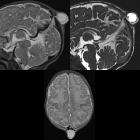
Newborn with
a bump on the back of the head. Sagittal T2 (above left) and sagital CISS (above right) MRI without contrast of the brain shows a fluid filled parietal lesion that contains meninges and that is connected by a short stalk to the dura. A persistent falcine sinus is present. Axial T2 MRI (below) shows the lesion to be in the midline.The diagnosis was atretic cephpalocele.
Atretic parietal cephaloceles (APC), also known as atretic cephaloceles, are small subscalp lesions that consist of dura, fibrous tissue, and dysplastic brain tissue.
Epidemiology
Common presentation in infants and young children.
Clinical presentation
Palpable midline parietal soft tissue mass.
Pathology
It is thought to represent involuted true cephalocele (meningocele or encephalocele) connected to dura mater via a fibrous stalk.
Associations
Increased incidence of intracranial anomalies.
Radiographic features
- subgaleal soft tissue mass with an intracranial extension via a sharply demarcated calvarial defect (cranium bifidum)
- CSF tract and vertical falcine vein point to the subcutaneous scalp mass
- vertically oriented primitive falcine vein
- fibrous stalk connecting the cephalocele
- focal fenestration of superior sagittal sinus at the atretic parietal cephalocele
- prominence of the superior cerebellar cistern and suprapineal recess
- superior peaking of the posterior tentorium
- spinning top configuration of the tentorial incisura
Differential diagnosis
Imaging differential considerations include:
- sinus pericranii
- dermoid or epidermoid cyst
- cephalohematoma
- sebaceous cyst
- vascular lesions (hemangioma)
Treatment and prognosis
The prognosis of atretic cephaloceles is generally good.
See also
Siehe auch:


 Assoziationen und Differentialdiagnosen zu atretische parietale Cephalocele: