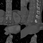
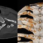
butterfly type vertebrae


Preschooler
with a faun’s tail in the midline of the back at the thoraco-lumbar junction. AP radiograph of the thoracolumbar spine shows a bone spur in the midline of the spinal canal at the T12-L1 level.The diagnosis was diastematomyelia.

Spinal cord
injury in an adult patient with thoracic butterfly vertebra: a case report and review of the literature. Magnetic resonance imaging (MRI) of the spine after injury. T1-weighted MRI (a) shows normalized signal intensities of the spinal cord. T2-weighted (b) and fat-suppressed MRI sequences (c) show a high-signal lesion in the spinal cord adjacent to the T11 vertebra. A coronal image (d) shows the presence of a butterfly vertebra at the T11 level. There was no signal change in the vertebral bodies or intervertebral discs

Spinal cord
injury in an adult patient with thoracic butterfly vertebra: a case report and review of the literature. Plain radiograph of the thoracic vertebrae (a, lateral view) shows anterior wedging of T11, which could be easily confused for a compression fracture. An anteroposterior view (b) shows a symmetrical defect with corticated margins in the T11 vertebral body

Butterfly
vertebra. Axial CT image demonstrates the sagittal cleft in the vertebral body


Butterfly
vertebra. Coronal CT imaging shows a sagittal cleft in T6.

Diastematomyelia
• Diastematomyelia - type II - Ganzer Fall bei Radiopaedia

Vertebral
anomalies • Butterfly vertebra - Ganzer Fall bei Radiopaedia

Butterfly
vertebra • Butterfly vertebra - Ganzer Fall bei Radiopaedia

Schmetterlingswirbel
(erster Sakralwirbel) in der Magnetresonanztomographie. Man erkennt den vertikalen Spalt durch den Wirbelkörper signalarm, also bindegewebig.

Schmetterlingswirbel
(erster Sakralwirbel) in der Magnetresonanztomographie. Man erkennt den vertikalen Spalt durch den Wirbelkörper signalarm, also bindegewebig.

Butterfly
vertebra • Butterfly vertebrae with kyphoscoliosis - Ganzer Fall bei Radiopaedia


Klippel-Feil
Syndrome with associated meningocoele and Sprengel deformity. Butterfly vertebra with elevation of the right scapula compared to left scapula

Butterfly
vertebra • Butterfly vertebra - Ganzer Fall bei Radiopaedia

Schmetterlingswirbel
(erster Sakralwirbel) in der Magnetresonanztomographie. Man erkennt den vertikalen Spalt durch den Wirbelkörper signalarm, also bindegewebig.

Butterfly
vertebra • Butterfly vertebra - Ganzer Fall bei Radiopaedia

Butterfly
vertebra • Lumbarized S1 butterfly vertebra - Ganzer Fall bei Radiopaedia

Butterfly
vertebra • Butterfly vertebra and anal atresia - Ganzer Fall bei Radiopaedia

Butterfly
vertebra • Butterfly and lumbosacral transitional vertebrae - Ganzer Fall bei Radiopaedia

Butterfly
vertebra • Butterfly vertebra - Ganzer Fall bei Radiopaedia

Butterfly
vertebra • Tracheo-esophageal fistula / esophageal atresia and clavicle fracture - Ganzer Fall bei Radiopaedia

Butterfly
vertebra • Cirrhosis - Ganzer Fall bei Radiopaedia

Schmetterlingswirbel.
Die Fehlbildung selbst ist rot eingefärbt - zum Vergleich: normaler Wirbel blau. Grün: Deformierung des Nachbarwirbels.

Butterfly
vertebra • Butterfly (photo) - Ganzer Fall bei Radiopaedia
Butterfly vertebra is a type of vertebral anomaly that results from the failure of fusion of the lateral halves of the vertebral body because of persistent notochordal tissue between them.
Pathology
Associations
- anterior spina bifida +/- anterior meningocele
- can be part of the Alagille syndrome
- Jarcho-Levin syndrome
- VACTERL association
Radiographic features
Plain radiograph
Widening of the affected vertebral body. The bodies above and below the butterfly vertebra adapt to the altered intervertebral discs on either side by showing concavities along the adjacent endplates.
Some bone bridging may occur across the defect which usually happens in the thoracic or lumbar segments of the spine.
Siehe auch:
- persistierende Chorda dorsalis
- angeborene Wirbelanomalien
- Diastematomyelie
- koronare Wirbelspalten
- VATER association
- Alagille-Syndrom
und weiter:

 Assoziationen und Differentialdiagnosen zu Schmetterlingswirbel:


