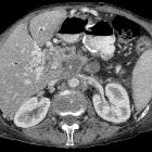
Celiac plexus block


Celiac plexus block under image guidance is an easy and safe percutaneous procedure with good outcomes for pain palliation in patients who have chronic abdominal pain related to the celiac ganglia.
This usually includes patients with advanced cancers, especially from upper abdominal viscera, such as the pancreas, stomach, duodenum, proximal small bowel, liver and the biliary tract, or due to enlarged lymph nodes.
History
Splanchnic nerve block for pain from the upper abdominal viscera was first described by Maxi Kappis in 1914 using bony landmarks from a posterior approach. He demonstrated that this could be used as a form of surgical anesthesia.
As image guidance started becoming widespread in the 1950s, Jones (1957) described the use of ethanol-induced celiac plexus neurolysis for long term pain relief. This method is now well-established.
Indications
- persistent, intractable abdominal or localized back pain due to malignant disease in the upper abdomen
- failure of standard pain control therapy
Contraindications
These are relative and include:
- severe uncorrectable coagulopathy or thrombocytopenia
- abdominal aortic aneurysm
- eccentric origin of the celiac artery
- inability to visualize local anatomy due to large overlying soft tissue mass
Procedure
Equipment
Although fluoroscopy was the earliest method of image guidance used, CT is the commonest modality that has been subsequently described to date. Some operators have described the use of ultrasound which allows for easy visualization of the celiac vessels but is operator and patient-dependent.
Technique
Celiac ganglia are located anterior to the crura of the diaphragm, over the anterolateral wall of the aorta bilaterally, and just caudal to the level of the origin of the celiac artery. Both anterior and posterior approaches may be used to access these, depending on the operator’s preferences and the safest route of access.
The anterior approach carries a reduced risk of neurologic complications since the needle tip is anterior to the spinal arteries and spinal canal. This also allows for a single puncture, reduced procedure time and use of a smaller volume of neurolytic agent. It also avoids the risk of puncturing the aorta and permits the patients to remain supine during the whole procedure.
20 to 50 mL of ethanol, with concentrations of 50–100%, is the most commonly used neurolytic agent in clinical practice. Phenol is also used as a neurolytic agent. Bupivacaine or lidocaine, as local anesthetics, have been used for celiac plexus block.
Postprocedural care
Patients are usually admitted for close hemodynamic and neurological monitoring overnight. They should be kept well-hydrated using IV fluids as necessary as there is a risk of periprocedural hypotension.
Complications
Pain
Local posterior abdominal and back pain during or immediately after a celiac plexus block has been reported commonly because of the ablative effect of the neurolytic agent.
Diarrhea
Often self-limiting, diarrhea occurs due to sympathetic blockade and unopposed parasympathetic efferent influence after the block. It usually resolves over approximately 48 hours.
Orthostatic hypotension
This may occur due to loss of sympathetic tone and dilated abdominal vasculature. It is usually transient (few hours) and can be managed conservatively with IV fluids as required.
Neurological
Paraplegia, leg weakness, sensory deficits, and paresthesias have rarely been reported. This is attributed to either direct injury of the spinal cord during the procedure or injection into the anterior spinal artery, which supplies the lower two thirds of the spinal cord .
Other
Puncture complications to the upper abdominal viscera (e.g. liver, stomach, pancreas and bowel) are rare.
Other rarely reported complications include impotence, gastroparesis, superior mesenteric vein thrombosis, chylothorax, pneumothorax, chemical pericarditis, aortic pseudoaneurysm, aortic dissection, hemorrhage and retroperitoneal fibrosis.
Outcomes
All patients should be interviewed before the procedure to obtain a baseline pain score to compare with post-procedural pain. A visual analog scale can be used to quantify a patient's subjective pain while the doses of pain medication taken can be used as objective markers of procedural outcome.
Siehe auch:
- Pneumothorax
- Pankreaskarzinom
- Chylothorax
- Arteria spinalis anterior
- Truncus coeliacus
- Aortendissektion
- Ganglion coeliacum
- retroperitoneale Fibrose allgemein
und weiter:

 Assoziationen und Differentialdiagnosen zu Zöliakusblockade:


