developmental dysplasia of the hip


Developmental dysplasia of the hip (DDH), or in older texts congenital dislocation of the hip (CDH), denotes aberrant development of the hip joint and results from an abnormal relationship of the femoral head to the acetabulum.
Unlike CDH, developmental dysplasia of the hip is not confined to congenital malformations and includes perturbations in development . There is a clear female predominance, and it usually occurs from ligamentous laxity and abnormal position in utero. Therefore, it is more common with oligohydramniotic pregnancies. This article describes the commonly used radiographic measurements and lines involved in DDH.
Epidemiology
The reported incidence of developmental dysplasia of the hip varies between 1.5 and 20 per 1000 births , with the majority (60-80%) of abnormal hips resolving spontaneously within 2-8 weeks (so-called immature hip).
Risk factors include :
- female gender (M: F ratio ~1:8)
- firstborn baby
- family history
- breech presentation
- oligohydramnios
- metatarsus adductus
- spina bifida
Clinical presentation
Developmental dysplasia of the hip is usually suspected in the early neonatal period due to the widespread adoption of clinical examination (including the Ortolani test, Barlow maneuvers, and Galeazzi sign). The diagnosis is then usually confirmed with ultrasound, although the role of ultrasound in screening is controversial .
Pathology
In general, the dysplastic hip has a ridge (neolimbus) in the superolateral region of the acetabulum composed hypertrophied fibrocartilage as a result of the abnormal joint congruity . In addition, there is very cellular hyaline cartilage allowing the femoral head to glide out of the acetabulum generating the palpable clunk known as the Ortolani sign .
Radiographic features
For imaging assessment of developmental dysplasia of the hip, ultrasound is the modality of choice prior to the ossification of the proximal femoral epiphysis. Once there is a significant ossification then an x-ray examination is required.
For some reason, the left hip is said to be more frequently affected . One-third of cases are affected bilaterally .
Ultrasound
Ultrasound is the test of choice in the infant (<6 months) as the proximal femoral epiphysis has not yet significantly ossified. Additionally, it has the advantage of being a real-time dynamic examination allowing the stability of the hip to be assessed with stress views.
Some values are used to 'objectively' assess morphology.
See: sonographic classification of developmental hip dysplasia
Alpha angle
The alpha angle is formed by the acetabular roof to the vertical cortex of the ilium. This is similar measurement as that of the acetabular angle (see below). The normal value is greater than or equal to 60º.
Beta angle
The beta angle is formed by the vertical cortex of the ilium and the triangular labral fibrocartilage (echogenic triangle). The normal value is less than 77º but is only useful in assessing immature hips when combined with the alpha angle.
Bony coverage (d:D ratio)
The percentage of the femoral epiphysis covered by the acetabular roof. A value of >50% is considered normal .
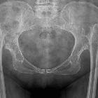
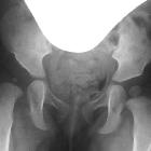
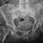
Plain radiograph
The key to plain film assessment of developmental dysplasia of the hip is looking for symmetry and defining the relationship of the proximal femur to the developing pelvis. The ossification of the superior femoral epiphyses should be symmetric. Delay of ossification is a sign of developmental dysplasia of the hip.
Hilgenreiner line
The Hilgenreiner line is drawn horizontally through the inferior aspect of both triradiate cartilages. It should be horizontal but is mainly used as a reference for the Perkin line and measurement of the acetabular angle.
Perkin line
The Perkin line is drawn perpendicular to Hilgenreiner line, intersecting the lateral most aspect of the acetabular roof. The upper femoral epiphysis should be seen in the inferomedial quadrant (i.e. below Hilgenreiner line, and medial to Perkin line)
Acetabular angle
The acetabular angle is formed by the intersection between a line drawn tangential to the acetabular roof and Hilgenreiner line, forming an acute angle. It should be approximately 30° at birth and progressively reduce with the maturation of the joint.
Shenton line
The Shenton line is drawn along the inferior border of the superior pubic ramus and should continue laterally along the inferomedial aspect of the proximal femur as a smooth line. If there is a superolateral migration of the proximal femur due to developmental dysplasia of the hip then this line will be discontinuous.
Extrusion index
The extrusion index is a percentage measure of bony coverage of the femoral head by acetabulum in patients with fully matured femoral epiphyses. A value of less than 25% has been reported as normal . The greater the degree of acetabular dysplasia, the greater the extrusion index.
Treatment and prognosis
Management options include:
- Pavlik harness: usually for younger patients (less than six months of age)
- closed reduction: usually for older patients
- open reduction (ORIF): much older patients or if closed reduction is not successful
Siehe auch:
- Shenton's line
- Perkin's line
- reimers index
- Hilgenreiner's line
- Oligohydramnion
- CE Winkel
- acetabular angle
- alpha angle (DDH)
- Sonographie des kindlichen Hüftgelenks
- cross-over sign Hüfte
- Angeborene Hüftluxation
- Extrusion Hüfte
- beta angle (DDH)
- Tragflächenwinkel
- Tiefe-zu-Weite-Index nach Heyman und Herndorn
und weiter:


 Assoziationen und Differentialdiagnosen zu Hüftdysplasie:

