epidurale intraspinale Raumforderung

Epidurale
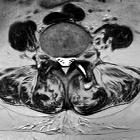
Lipomatose, die in Kombination mit multiplen degenerativ bedingten Hypertrophien und einem Discusprolaps (L4/5 nach oben sequestriert) zu einer spinalen Stenose führt. Bezeichnend ist die Einengung des Duralsackes nicht nur auf Höhe der Bandscheiben, sondern auch auf Höhe der Wirbelkörper, wie in dem axialen T2w-Schnitt im rechten Bild (kein Liquorsaum mehr). Eine längere Glukokortikoid-Einnahme war bekannt und durchaus als Ursache zu verdächtigen, zumal eine Voruntersuchung vor der Medikation noch nicht dieses Bild zeigte.

Zufallsbefund
einer lateralen meningealen Zyste auf Höhe BWK 12. (MRT wegen LWK-4-Fraktur). Die Zyste findet sich auf Höhe des Wirbelkörpers im Gegensatz zu einer Wurzeltaschenzyste, die sich in die Foramina ausdehnt und diese ggf. aufweitet. Die in T2 signalreichere Darstellung ggü. dem Liquor im Duralsack ist durch Pulsationen in letzterem erklärt.

Facettengelenksarthrose
mit Facettengelenks-Zyste von rechts nach intraspinal mit hochgradiger spinaler Enge.

Intraspinale,
epidurale, eingeblutete Synovialzyste Facettengelenk mit akuter Klinik mit Schmerz und Kaudasymptomatik. Oben sagittal T1 nativ (hell !), T2, STIR, unten T2 axial , T1 KM FS axial und sagittal.

Hirayama
disease • Hirayama disease - Ganzer Fall bei Radiopaedia

Epidural
spinal cord compression as initial clinical presentation of an acute myeloid leukaemia: case report and literature review. Spine MRI showing spinal cord compression at T4-T9 level by posterior epidural mass. a Sagittal view showing a T4T9 posterior mass which is isointense on T2 weighted sequence. b Sagittal view T1 sequence showing a hypointense T4T9 posterior lésion well demarcated from the dura. c Sagittal view showing a contrast enhancement of the T4-T9 lesion. d Axial view: The lesion is hypointense on T1 weighted sequence. e Axial view: The lesion is isointense on T2 sequence at the level of T5. f Axial view showing the enhancing lesion at the level of T5 wth a good demarcation from the dura

Facettengelenksarthrose
mit Facettengelenks-Zyste nach dorsal. Es findet sich vermehrt Flüssigkeit im Gelenkspalt, links mehr als rechts. Die Zyste schließt sich dorsal links an.

Epidural
metastatic melanoma. Subtraction images before and after contrast show a marked enhancement of a tail-like structure adjacent to the lesion, superior and inferior of the lesion (green arrows).

Spinal
arachnoid cyst • Lumbar epidural arachnoid cyst - Ganzer Fall bei Radiopaedia

Spinal
epidural abscess • Epidural abscess of the cervical spine - Ganzer Fall bei Radiopaedia

Spinal
epidural mass • Spinal epidural hematoma - Ganzer Fall bei Radiopaedia

Extramedullary
hematopoiesis • Extramedullary hematopoiesis - spinal epidural lesions - Ganzer Fall bei Radiopaedia

Spinal
epidural mass • Extradural thoracic spinal cavernous malformation - Ganzer Fall bei Radiopaedia

Spinal
epidural mass • Spinal extradural Ewing sarcoma - Ganzer Fall bei Radiopaedia
The differential diagnosis for a spinal epidural mass includes:
- epidural metastasis
- epidural abscess
- herniated nucleus pulposus
- epidural hematoma
- epidural arteriovenous malformation
- epidural angiolipoma
- epidural lipomatosis
Siehe auch:
- epidurale Lipomatose
- extramedulläre Hämatopoese
- epidurales Hämatom
- Synovialzysten der Facettengelenke
- intraspinale epidurale zystische Raumforderungen
- extraossäres Plasmozytom
- intraspinale Tumoren
- eingeblutete Synovialzyste der Facettengelenke
- intraspinal extramedullary haematopoiesis
- angiolipoma thoracic spine
- epidurales spinales Myelosarkom
- spinaler Epiduralabszess
- Hirayama-Krankheit
- spinal angiomyolipoma
- spinale epidurale Metastasen
- spinale epidurale Kontrastmittelaufnahme
- Migration eines Bandscheibensequesters
- intraspinales epidurales Angiolipom
- spinales epidurales Hämatom
und weiter:

 Assoziationen und Differentialdiagnosen zu epidurale intraspinale Raumforderung:
spinale
epidurale Kontrastmittelaufnahme


