exudative Retinitis


Exudative retinitis, also known as retinal telangiectasis or Coats disease, is a rare congenital disease affecting the eyes and is a cause of leukocoria.
Epidemiology
It occurs predominantly in young males, with the onset of symptoms generally appearing in the first decade of life with a peak age of onset between 3-9 years of age . Although the condition is also recognized in adults, in which case it is usually less severe, the vast majority are diagnosed before the age of 20 years . There is well recognized male predominance (69-85%) .
Clinical presentation
Clinical presentation is usually with leukocoria or strabismus, although eventually, complete blindness may develop. The diagnosis is usually made with indirect ophthalmoscopy . The disease is unilateral in 80-90% of cases .
Associations
Pathology
The exact etiology is unknown and the disease is a non-hereditary disorder. The disorder is primarily one of the weak capillaries of the retina, resulting in progressive retinal detachment due to an exudative sub-retinal collection.
These exudates contain cholesterol crystals, lipid-laden macrophages and a few red cells. However, hemosiderin is minimal .
Radiographic features
Ultrasound
Ultrasound is an excellent modality for the assessment of the globe, however, is hampered in advanced cases by limited specificity and difficulty in distinguishing Coats disease from non-calcified retinoblastoma .
CT
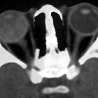
Findings depend on the stage of the disease. Early on, the examination may be normal. Enhancement may be seen at the margins of the exudate and may have a V-shaped pattern similar to retinal detachment . In advanced cases the affected globe is hyperdense due to proteinaceous exudates and the vitreous space may be obliterated due to extensive retinal detachment. Calcification is uncommon but has been reported .
The affected eye in Coats disease is usually significantly smaller than the non-affected eye in older children and is thought to represent an impairment of growth rather than the reduction in the volume of a previously normal globe .
MRI
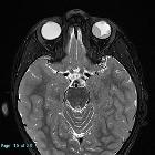
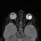
MRI appearances are similar to CT with better contrast resolution and the ability to visualize fainter enhancement, but reduced ability to visualize calcium. MRI is also able to visualize globe size.
- T1: high signal due to proteinaceous nature of the exudates
- T2: high signal due to proteinaceous nature of the exudates
- T1 C+ (Gd)
- enhancement of the detached retina may be visible
- rarely an 'enhancing mass' may appear to be present in advanced cases
History and etymology
This disease is named after a Scottish ophthalmologist George Coats (1876-1915) who first described it in 1908 .
Treatment
Treatment remains controversial, and options include laser photocoagulation, cryotherapy and surgical repair of retinal detachment.
In general, patients who present at an earlier age will fare worse than older patients .
Differential diagnosis
- persistent hyperplastic primary vitreous (PHPV)
- retinopathy of prematurity
- non-calcifying retinoblastoma
- this is especially worrisome in younger patients
- CT and MRI with contrast should demonstrate a mass
- the globe is normal or increased in size
- 95% of retinoblastomas are calcified
Siehe auch:
- Retinoblastom
- Leukokorie
- Netzhautablösung
- persistierender hyperplastischer primärer Glaskörper (PHPV)
- Retinopathia praematurorum
- VATER association
und weiter:

 Assoziationen und Differentialdiagnosen zu Morbus Coats:


