generalized increased bone density in adults


Osteopetrosis.
ADO: lateral spine radiograph, age 4 years. Note sclerosis of vertebral endplates (arrows) resulting in "sandwich vertebrae" appearance.

Paget disease
(bone) • Paget disease of the skull - Ganzer Fall bei Radiopaedia


Myelodysplastisches
Syndrom mit Myelosklerose: links T1, rechts T2. Beim MDS würde man eigentlich ein niedriges T1-Signal und ein hohes T2-Signal erwarten. Das hier auch niedrige T2-Signal legt den Verdacht einer begleitenden Myelosklerose nahe.

Generalized
increased bone density in adults • Systemic mastocytosis - Ganzer Fall bei Radiopaedia

Systemic
mastocytosis. Abdominal X-ray showed diffuse sclerosis of the lumbar vertebrae, sacrum, iliac and femoral heads.

Thoracic
myelopathy caused by ossification of ligamentum flavum of which fluorosis as an etiology factor. Anteroposterior view radiograph of both forearms showed significant calcifications of interosseous membranes of forearm.

Osteopoikilosis.
Shoulder joint radiographs detected elliptic and longitudinal sclerotic bone lesions in epiphyseal and metaphyseal region of proximal humeral bones, distal clavicles and scapulae.

Osteopoikilosis.
Radiographic findings of KL grade 3 osteoarthritis of hip joints associated with multiple, scattered, juxta-articular and metaphyseal, ovoid and longitudinal radiopacities.[8]

Osteopetrosis.
ADO: Radiograph of left femur, age 4 years. Note Erlenmeyer flask deformity of distal femur (arrows) and generalised increased bone density.

Osteopetrosis.
Pycnodysostosis: Lateral skull radiograph, age 3 years. Note loss of the mandibular angle (arrow) and increased thickness of vault.

Osteopetrosis.
Pycnodysostosis: Hand radiograph, age 3 years. Note acro-osteolysis in the distal phalanx of thumb and index fingers (arrows) and generalised increased bone density.

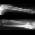
Infant with a
newly diagnosed adrenal mass. AP radiographs of the humerus (above) and radius and ulna (below) show lesions involving the proximal humeral diaphysis, entire radial diaphysis and proximal ulna diaphysis all of which are lytic in appearance and having a wide zone of transition and associated faint periosteal reaction.The diagnosis was bone metastases from neuroblastoma.

Preschooler
with short stature. AP and lateral radiographs of the skull (above) and axial CT without contrast of the brain (below) shows diffusely sclerotic skull bones with a thickened diploic space and diastatis of the coronal and lambdoid and sagittal sutures.The diagnosis was pyknodysostosis.

Generalized
increased bone density in adults • Osteoblastic metastases (prostate carcinoma) - Ganzer Fall bei Radiopaedia

Systemic
mastocytosis. CT scan shows the same findings already noted previously at the plain radiographic studies: diffuse bone sclerosis.

Osteopoikilosis.
Coronal and axial MDCT findings of the pelvis showed symmetric, mixed lytic and hyperdense ovoid and irregular sclerotic lesions scattered in peri-acetabular regions and in epiphyseal and metaphyseal parts of the proximal femora.

Systemic
mastocytosis. Sagittal CT scan MPR reformation showing diffuse vertebral sclerosis.

Thoracic
myelopathy caused by ossification of ligamentum flavum of which fluorosis as an etiology factor. a, b. T1 and T2 weight MRI of thoracic spine showed continuous multi-level ossification of ligamentum flavum between T7–12. c. CT scan showed ossified ligamentum flavum, note that there was a thin gap between the ossified ligament and the lamina.

Generalized
increased bone density in adults • Renal osteodystrophy and brown tumors - Ganzer Fall bei Radiopaedia

Generalized
increased bone density in adults • Diffuse bone metastases - prostate cancer - Ganzer Fall bei Radiopaedia

Generalized
increased bone density in adults • Renal osteodystrophy - Ganzer Fall bei Radiopaedia

Generalized
increased bone density in adults • Breast cancer metastasis - Ganzer Fall bei Radiopaedia

The causes of generalized increase in bone density in adult patients, also known as generalized or diffuse osteosclerosis, can be divided according to broad categories:
- hematological disorders
- myelosclerosis
- marrow cavity is narrowed by endosteal new bone
- patchy lucencies due to the persistence of fibrous tissue (generalized osteopenia in the early stages due to myelofibrosis)
- hepatosplenomegaly
- sickle cell disease
- osteosclerosing multiple myeloma
- myelosclerosis
- metabolic
- poisoning
- fluorosis
- with periosteal reaction, prominent muscle attachments and calcification of ligaments and interosseous membranes
- changes are most marked in the innominate bones and lumbar spine
- fluorosis
- neoplastic (more commonly multifocal than generalized)
- malignancy
- osteoblastic metastases: most commonly prostate and breasts
- lymphoma: infiltrative
- leukemia: infiltrative
- mastocytosis
- sclerosis of marrow-containing skeleton with patchy areas of radiolucency
- urticaria pigmentosa
- can have symptoms and signs of carcinoid syndrome
- idiopathic (more commonly multifocal than generalized)
- Paget disease: coarsened trabeculae, bony expansion and thickened cortex
- congenital: sclerosing bone dysplasias
- other
- hepatitis C associated osteosclerosis (HCAO)
There are several mnemonics for dense bones.
See also
Siehe auch:
- osteoblastische Knochenmetastasen
- diffuse bony sclerosis (mnemonic)
- Morbus Paget des Knochens
- Osteopoikilose
- Sichelzellenanämie
- Osteopetrose
- Osteomyelofibrose
- Renale Osteodystrophie
- Adenokarzinom der Prostata
- Pyknodysostose
- Melorheostose
- Fluorose
- Osteopathia striata
- Mastozytose
- osteomesopyknosis
- Leukämie
- Skeletaler Lymphombefall
- differential diagnosis of generalised increased bone density in adults
- sclerosing bony dysplasias
und weiter:

 Assoziationen und Differentialdiagnosen zu diffuse skelettale Sklerosierung:


