Globus pallidus


Insular
cortex • Basal ganglia location - Gray's anatomy illustration - Ganzer Fall bei Radiopaedia

Basal ganglia
• Brainstem nuclei and their connections - Gray's anatomy illustration - Ganzer Fall bei Radiopaedia

Basal ganglia
• Organisational structure of basal ganglia - Ganzer Fall bei Radiopaedia

Basal ganglia
• Basal ganglia (Gray's illustrations) - Ganzer Fall bei Radiopaedia

Corpus
striatum • Corpus striatum (Gray's illustration) - Ganzer Fall bei Radiopaedia
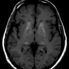
The globi pallidi (singular: globus pallidus) are paired structures and one of the nuclei that make up the basal ganglia. Each globus pallidus is a subcortical structure at the base of the forebrain and in anatomical relation to the caudate nucleus and putamen. It forms the lentiform nucleus with the putamen.
Each globus pallidus is itself subdivided into two parts, the globus pallidus internus and globus pallidus externus, separated by an internal medullary lamina.
Developmentally, the globus pallidus is a part of the diencephalon that has migrated to the telencephalon, but it is still considered to be part of the basal ganglia.
Related pathology
See also
- basal ganglia T1 hypointensity
- basal ganglia T1 hyperintensity
- basal ganglia T2 hypointensity
- basal ganglia T2 hyperintensity
Siehe auch:
- Basalganglienverkalkungen
- T2 hyperintense Basalganglien
- basal ganglia T1 hyperintensity
- Putamen
- Basalganglien
- diencephalon
- Nucleus lentiformis
- Nucleus caudatus
- decreased T2 signal in the basal ganglia
und weiter:

 Assoziationen und Differentialdiagnosen zu Globus pallidus:


