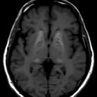
basal ganglia T1 hyperintensity

Basal ganglia
T1 hyperintensity • Bilateral basal ganglia and thalamic T1 hyperintensities - Ganzer Fall bei Radiopaedia

Basal ganglia
T1 hyperintensity • Non-ketotic hyperglycemic hemichorea - Ganzer Fall bei Radiopaedia

Basal ganglia
T1 hyperintensity • Reversible basal ganglia T1 hyperintensity - Ganzer Fall bei Radiopaedia

Basal ganglia
T1 hyperintensity • Acquired hepatocerebral degeneration - Ganzer Fall bei Radiopaedia

T1-hypointense
Stammganglien am ehesten als Ausdruck einer hepatischen Enzephalopathie. Nebenbefundlich betonte perivaskuläre Räume.
There are many causes of basal ganglia T1 hyperintensity, but the majority relate to deposition of T1-intense elements within the basal ganglia such as:
- calcium
- idiopathic calcification
- calcium and phosphate abnormalities
- hepatic failure
- acquired non-wilsonian hepatocerebral degeneration
- Wilson disease (copper)
- hepatic encephalopathy
- toxins/ischemia
- carbon monoxide (usually low T1 signal; unless associated with hemorrhage)
- hyperalimentation or long term parenteral nutrition (manganese)
- hyperglycemia-associated choreaballism : non-ketotic hyperglycemic hemichorea (NHH)
- previous administration of linear gadolinium chelates
- global hypoxia
- blood
- methemoglobin in intracranial hemorrhage
- hemorrhagic infarct
- Japanese encephalitis
- hamartoma in neurofibromatosis type 1
See also
Siehe auch:
- Basalganglienverkalkungen
- Neurofibromatose Typ 1
- T2 hyperintense Basalganglien
- Morbus Wilson
- Basalganglien
- ADC abnormality of the basal ganglia
- acquired hepatocerebral degeneration
- eye of tiger sign
- Hepatische Enzephalopathie
- decreased T1 signal in the basal ganglia
- decreased T2 signal in the basal ganglia
- basal ganglia signal abnormalities
- diabetische Striatopathie
und weiter:

 Assoziationen und Differentialdiagnosen zu basal ganglia T1 hyperintensity:
ADC
abnormality of the basal ganglia


