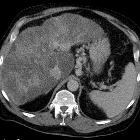
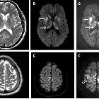
restricted diffusion in the basal ganglia

Basal ganglia
restricted diffusion • Artery of Percheron territory infarct - Ganzer Fall bei Radiopaedia

Basal ganglia
restricted diffusion • Rabies encephalomyelitis - Ganzer Fall bei Radiopaedia

Basal ganglia
restricted diffusion • Creutzfeldt Jakob disease - Ganzer Fall bei Radiopaedia

Basal ganglia
restricted diffusion • Japanese encephalitis - Ganzer Fall bei Radiopaedia

Basal ganglia
restricted diffusion • Hypoglycemic encephalopathy - Ganzer Fall bei Radiopaedia
Restricted diffusion in the basal ganglia (and thalami) is seen in a variety of conditions, often associated with other signal abnormality of the basal ganglia. Care should be taken to confirm that increased signal on DWI actually represents restricted diffusion rather than merely T2 shine through.
It is important to note that each entity listed below has a variable pattern of involvement of the deep grey matter structures (basal ganglia and thalami).
Causes include :
- acute exposure to respiratory chain metabolic toxins
- Wilson disease (early)
- hyperammonemia
- hypoglycemia
- hypoxic ischemic encephalopathy
- osmotic demyelination
- Wernicke encephalopathy
- Creutzfeldt-Jakob disease
- deep cerebral venous thrombosis
- arterial infarction
- top of basilar syndrome
- perforator infarct (usually unilateral)
- artery of Percheron infarct
- flavivirus encephalitis
See also
Siehe auch:
- Leberzirrhose
- Korsakow-Syndrom
- Zentrale pontine Myelinolyse
- T2 hyperintense Basalganglien
- Morbus Wilson
- Creutzfeldt-Jakob-Krankheit
- Kohlenmonoxidintoxikation
- diffusionsgewichtete Bildgebung
- decreased T1 signal in the basal ganglia
- decreased T2 signal in the basal ganglia
- basal ganglia signal abnormalities
- respiratory chain metabolic toxins
- Enzephalitis durch Flaviviren
- percheron artery infarct
- top of the basilar artery syndrome
- signal abnormality of the basal ganglia
- t2 shine through
- increased T1 signal in the basal ganglia
und weiter:

 Assoziationen und Differentialdiagnosen zu ADC abnormality of the basal ganglia:


