basal ganglia


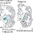
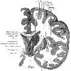


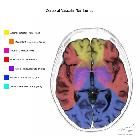
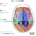
The basal ganglia are a group of grey matter nuclei in the deep aspects of the brain that is interconnected with the cerebral cortex, thalami and brainstem.
In a strict anatomical sense, it contains three paired nuclei that together comprise the corpus striatum:
Functionally, two additional nuclei are also part of the basal ganglia:
Terminology
Whilst very widely used in English, the term 'basal ganglia' is actually a misnomer, as a ganglion is a collection of nerve cell bodies outside of the central nervous system. The equivalent within the central nervous system is termed 'nucleus', as reflected in the official term for the basal ganglia in the Terminologia Anatomica, 'nuclei basales', the English translation of which is 'basal nuclei'.
This is also illustrated by the name of each individual basal nucleus, e.g. caudate nucleus, lentiform nucleus, subthalamic nuclei, etc.
Radiographic features
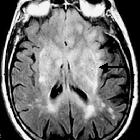
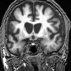
The basal ganglia are normally isodense/isointense to the cortex. Because the globus pallidus has more myelin content compared with the putamen, it usually appears slightly more hypointense on T2WI, GRE, and SWI images. Age-related calcium deposition in the globus pallidus initially results in increased T1 signal intensity and subsequently, when calcification exceeds 40%, signal loss in all sequences. Aging with consequent iron deposition in the putamen results in a gradual decrease of T2/T2*/SWI signal intensity in the putamen. This is more pronounced in the 8 or 9 decade of life.
Related pathology
- basal ganglia calcification
- Parkinson disease
- Huntington disease
- Hallervorden-Spatz syndrome
- central pontine myelinolysis
- gliomatosis cerebri
- cerebral microhemorrhage
See also
Siehe auch:
- Gliomatosis cerebri
- Basalganglienverkalkungen
- Thalamus
- Zentrale pontine Myelinolyse
- Globus pallidus
- T2 hyperintense Basalganglien
- Putamen
- substantia nigra
- zerebrale Mikroblutungen
- Anatomie Hirnstamm
- ADC abnormality of the basal ganglia
- corpus striatum
- Nucleus lentiformis
- Neurodegeneration mit Eisenablagerung im Gehirn
- Chorea Huntington
- Nucleus subthalamicus
- Nucleus caudatus
- decreased T1 signal in the basal ganglia
- decreased T2 signal in the basal ganglia
- basal ganglia signal abnormalities
- increased T1 signal in the basal ganglia
und weiter:
- Arteria cerebri media
- basal ganglia T1 hyperintensity
- neuroradiologisches Curriculum
- Cerebrum
- extrapontine myelinolysis
- Subakute sklerosierende Panenzephalitis
- Claustrum
- extrapyramidal system
- kernicterus
- sub acute necrotizing encephalomyelopathy
- respiratory chain metabolic toxins
- eosinophilia myalgia syndrome
- sub acute sclerosing panencephalitis


 Assoziationen und Differentialdiagnosen zu Basalganglien: