extrapontine myelinolysis

Central
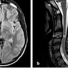
Pontine and Extra Pontine Myelinolysis. T2-FLAIR axial image showing symmetrical hyperintensities in bilateral caudate, lentiform nucleus and thalami, consistent with extra pontine myelinolysis.

Central
Pontine and Extra Pontine Myelinolysis. T2-FLAIR Coronal image showing symmetrical caudate, lentiform hyperintensities coupled with central pontine hyperintensities suggesting pontine and extra pontine myelinolysis.

Central
Pontine and Extra Pontine Myelinolysis. DW axial image showing hyperintensities in both caudate, both lentiform nucleus and in thalami. These are secondary to T2 shine through and are unlikely to be diffusion restriction (look at figure 5 for ADC image).
Extrapontine myelinolysis (EPM) is one of the complications occurring secondary to rapid correction of hyponatremia, and is, along with central pontine myelinolysis encompassed by the more recent term osmotic demyelination syndrome.
In the vast majority of cases it is associated with central pontine myelinolysis but it can also (rarely) occur as an isolated entity.
Pathology
Location
In extrapontine myelinolysis, sites of involvement include:
- ventrolateral thalami
- basal ganglia
- caudate nuclei
- internal capsule
- external capsule
- extreme capsule
- grey-white matter junction
- splenium of the corpus callosum
Siehe auch:
- Pons
- Encephalomyelitis disseminata
- Mesencephalon
- Astrozytom
- Basalganglien
- Anatomie Hirnstamm
- Demyelinisierende Erkrankung
- T2 hyperintense Ponsläsionen
- Pseudobulbärparalyse
- beam hardening phenomenon
und weiter:

 Assoziationen und Differentialdiagnosen zu extrapontine myelinolysis:


