central pontine myelinolysis

Osmotic
demyelination syndrome • Central pontine myelinolysis - Ganzer Fall bei Radiopaedia

Central
Pontine and Extra Pontine Myelinolysis. T2-FLAIR Coronal image showing symmetrical caudate, lentiform hyperintensities coupled with central pontine hyperintensities suggesting pontine and extra pontine myelinolysis.

Osmotic
demyelination syndrome • Central pontine myelinolysis - Ganzer Fall bei Radiopaedia

Trident sign
(osmotic demyelination) • Osmotic demyelination syndrome - Ganzer Fall bei Radiopaedia

Trident sign
(osmotic demyelination) • Osmotic demyelination syndrome - Ganzer Fall bei Radiopaedia

Trident sign
(osmotic demyelination) • Trident sign - osmotic demyelination - Ganzer Fall bei Radiopaedia

Osmotic
demyelination syndrome • Osmotic demyelination syndrome - Ganzer Fall bei Radiopaedia

Osmotic
demyelination syndrome • Osmotic demyelination syndrome - Ganzer Fall bei Radiopaedia

Osmotic
demyelination syndrome • Central pontine myelinolysis - Ganzer Fall bei Radiopaedia

Osmotic
demyelination syndrome • Central pontine myelinolysis - Ganzer Fall bei Radiopaedia

Central
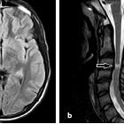
pontine myelinolysis during treatment of hyperglycemic hyperosmolar syndrome: a case report. Diffusion-weighted imaging of the brain. a Image 72 h after admission. b Image 30 days after admission. Arrowheads indicate high-intensity regions in the pons

Diffuse large
B-cell lymphoma presenting with central pontine myelinolysis: a case report. Brain magnetic resonance scan. The hyperintense lesion in the central pons in T2-weighted magnetic resonance imaging decreased and was almost undetectable after several courses of chemotherapy. (a) Before treatment, (b) after two courses, and (c) after seven courses of chemotherapy

Osmotic
demyelination syndrome • Osmotic demyelination syndrome - Ganzer Fall bei Radiopaedia

Osmotic
demyelination syndrome • Osmotic demyelination syndrome - Ganzer Fall bei Radiopaedia

Osmotic
demyelination syndrome • Central pontine and extrapontine myelinolysis - Ganzer Fall bei Radiopaedia

Osmotic
demyelination syndrome • Central pontine myelinolysis - Ganzer Fall bei Radiopaedia

Osmotic
demyelination syndrome • Central pontine myelinolysis - Ganzer Fall bei Radiopaedia

Plasma
exchange successfully treats central pontine myelinolysis after acute hypernatremia from intravenous sodium bicarbonate therapy. MRI of the brain revealed symmetric, high-intensity signal in the pons with sparing of the peripheral portion, suggesting central pontine myelinolysis (A, axial view and B, sagittal view).

Hyperglycemia-related
central pontine demyelinization after a binge-eating attack in a patient with type-2 diabetes: a case report. MRI image of the brain (a FLAIR cor) and cranial computed tomography image of the brain (b CE-cranial); arrows indicate the central pontine-demyelinization area

Hyperglycemia-related
central pontine demyelinization after a binge-eating attack in a patient with type-2 diabetes: a case report. MRI images of the brain (a and b: T2-weighted, c and d: B = 1000 DWI); arrows indicate the central pontine-demyelinization area

Central
Pontine and Extra Pontine Myelinolysis. T2-FLAIR axial image showing hyperintensity in the central pons suggesting pontine myelinolysis.
Central pontine myelinolysis (CPM) is now more commonly referred to as osmotic demyelination syndrome, which recognizes that the same phenomenon is also seen in other areas of the brain (previously known as extrapontine myelinolysis).
As such the condition is described in the osmotic demyelination syndrome article.
Siehe auch:
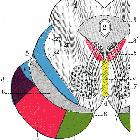
- Pons
- Mesencephalon
- Encephalomyelitis disseminata
- Anatomie Hirnstamm
- Basalganglien
- Demyelinisierende Erkrankung
- T2 hyperintense Ponsläsionen
- Alkoholinduzierte ZNS-Veränderungen
- Pseudobulbärparalyse
- Hyponatriämie
- pontine Tumoren
- Astrozytom des Pons
- beam hardening phenomenon
und weiter:


 Assoziationen und Differentialdiagnosen zu Zentrale pontine Myelinolyse: