inferior vena cava (IVC)


The inferior vena cava (IVC) drains venous blood from the lower trunk, abdomen, pelvis and lower limbs to the right atrium of the heart.
Gross anatomy
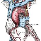
The inferior vena cava is formed by the confluence of the two common iliac veins at the L5 vertebral level. The IVC has a retroperitoneal course within the abdominal cavity. It runs along the right side of the vertebral column with the aorta lying laterally on the left. Various other veins drain into the IVC along its course before it passes through the diaphragm at the caval hiatus at the T8 level. Its intrathoracic course is very short before draining into the right atrium at the inferior cavoatrial junction.
Tributaries
- T8: paired inferior phrenic veins
- T8: hepatic veins
- L1: right suprarenal vein
- L1: renal veins
- L2: right gonadal vein
- L1-L5: lumbar veins
- L5: common iliac veins (origin)
Since the IVC is not a midline structure, there is a degree of asymmetry of drainage, e.g. the gonadal and suprarenal veins drain into the IVC on the right side, but into the left renal vein on the left.
Relations
- anterior: right common iliac artery, right gonadal vessels, third part of duodenum (D3), common bile duct, portal vein, head of pancreas, first part of duodenum (D1), epiploic foramen, liver
- posterior: lower lumbar vertebrae, intervertebral discs, anterior longitudinal ligament, right psoas major, sympathetic trunk, right celiac ganglion, right 3rd and 4th lumbar arteries, right renal artery, right suprarenal artery, right inferior phrenic arteries
- lateral (left): abdominal aorta, caudate lobe of the liver, right crus
- lateral (right): right kidney, right ureter, second part of duodenum (D2), liver
Development
Normal IVC has a complex embryological development with many embryological veins contributing to different parts:
- right vitelline vein: forms suprahepatic and hepatic segments of IVC
- right subcardinal vein: forms suprarenal segment
- right subsupracardinal anastomosis: forms renal segment
- right supracardinal vein: forms infrarenal segment
- right posterior cardinal vein: forms distal most IVC and its bifurcation into common iliac veins
Variant anatomy
Inferior caval abnormalities are typically the result of abnormal embryologic development involving the vitelline, posterior cardinal, subcardinal and supracardinal veins :
- absence of IVC (entire or only the infrarenal segment)
- IVC duplication
- azygos continuation of the IVC
- left sided IVC
- circumcaval ureter
- circumaortic venous collar
- IVC webs
- extrahepatic portocaval shunt (Abernethy malformation)
- marsupial cava
Rarely a Eustachian valve at the inferior cavoatrial junction may be present.
Related pathology
Siehe auch:
- Varianten der Vena cava inferior
- Zwerchfell
- Becken
- Vena cava superior
- Bauchhöhle
- Vena cava inferior Thrombose
und weiter:
- Echinococcus Leber
- retroperitoneal organs (mnemonic)
- Foramen epiploicum
- Aorta abdominalis
- vena caval foramen
- Snowman heart
- abdominal surface anatomy
- portal venous system
- ovaries
- Normale Herzkonfiguration im Röntgen-Thorax
- Nabelvenenkatheter
- totale Lungenvenenfehlmündung
- Arteria renalis
- great vessels
- retroperitoneales Leiomyosarkom
- Cavafilter
- accessory right inferior hepatic vein
- Ovar
- tension pneumoperitoneum
- fishhook ureters
- Vena cava
- Hoffman-Rigler sign
- Vena cava inferior Rechtsherzinsuffizienz
- gradman and Steinburg IVC aneurysm classification
- korrekte Position Nabelvenenkatheter


 Assoziationen und Differentialdiagnosen zu Vena cava inferior:


