ovaries


The ovaries are paired female gonads of the reproductive and endocrine systems. They lie within the ovarian fossa on the posterior wall of the true pelvis.
Gross anatomy
The ovaries are firm and ovoid in shape and measure approximately 1.5-3.0 cm × 1.5-3.0 cm × 1.0-2.0 cm (length x width x thickness) (corresponding to a volume of 1.2-9.4 cm). An ovary typically weighs 2-8 g, however, they change during life and double in size in pregnancy.
Typically they lie on the peritoneum of the pelvic wall in a shallow fossa in the angle between internal and external iliac vessels on the obturator nerve but have variable location secondary to their mobility. They are oriented with their long axis oblique, with lateral and medial surfaces, uterine and tubal ends, and mesovarian and free borders.
The suspensory ligament of the ovary, a peritoneal fold, runs from the sidewall of the pelvis to the ovary. The ovarian vessels run in this ligament, crossing over the external iliac vessels.
Each ovary is attached to the posterior lamina of the broad ligament by the mesovarium, which is continuous with its outer coat. A third attachment, the ovarian ligament, is a continuation of the round ligament and attaches the ovary to the side of the uterus.
Despite all its attachments, the ovary is very mobile, especially in women who have had children. It is frequently found behind the uterus in the pouch of Douglas and has a variable relationship with the uterus:
- anteflexed uterus: lateral or posterolateral
- retroflexed uterus: superolateral
Relations
- anteriorly: broad ligament, mesovarium, obliterated umbilical artery
- posteriorly: ureter, internal iliac vessels, suspensory ligament with ovarian vessels
- superiorly: external iliac vessels
- inferiorly: levator ani
- medially: ovarian ligament attaching ovaries to uterus, pararectal fossa and rectouterine pouch, bowel loops
- laterally: obturator vessels and nerves in ovarian fossa, obturator internus and fascia, parietal peritoneum of pelvic wall
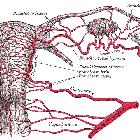
Arterial supply
The primary blood supply to the ovary is the ovarian artery, although there is some anastomosis with branches of the uterine artery
- ovarian artery
- ovarian branches from the uterine artery
Venous drainage
- pampiniform plexus drains into the ovarian veins
- right ovarian vein drains into the inferior vena cava
- left ovarian vein drains into the left renal vein
Lymphatic drainage
- drain alongside the ovarian vessels either to para-aortic nodes, or
- follow para-uterine vessels to iliac nodes, or
- alternative routes include inguinal nodes via round ligament or reaching the contralateral ovary by passing across the fundus of the uterus
Innervation
- sympathetic supply from T10 and T11 spinal segments via aortic plexus and its branches
- parasympathetic supply from inferior hypogastric plexus via uterine artery responsible for vasodilatation
- sensory fibers accompany sympathetic nerves
Variant anatomy
- suprapelvic position on psoas major
- inguinal canal location
- spheroidal, flattened, crescentic shape
- supernumerary ovaries
- absence or hypoplasia of one or both ovaries
- ectopic adrenal or thyroid tissue
Radiographic features
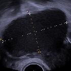
Ultrasound
- first-line investigation
- homogeneous echotexture with a central echogenic medulla
- volume on ultrasound can be calculated with the following formula :
- 0.523 × length (cm) × width (cm) × depth (cm)
- volume is expressed in cubic centimeters (cm)
CT
- can be difficult to identify or may be confused with enlarged lymph nodes
- its position may be determined by the following techniques :
- anterior or anterolateral to the pelvic ureter if in a typical position
- tracking the ovarian vein from the IVC or left renal artery anterior to the psoas major to the region where the ovary lies
- suspensory ligament of the ovary
- usually, extends from the ovary to the external or common iliac vessels; most easily identifiable pelvic ligament
- the ovary can be differentiated from lymph nodes, which are extraperitoneal and displace the ureter medially or anteromedially or efface the common iliac vessels when enlarged
Related pathology
- Müllerian duct abnormalities
- ectopic pregnancy: ovarian
- pelvic inflammatory disease
- endometriosis
- ovarian cyst
- ovarian dermoid
- ovarian cancer
- polycystic ovary
- polycystic ovarian syndrome
Siehe auch:
- Uterus
- Neoplasien des Ovars
- Endometriose
- Vena cava inferior
- Ovarialzyste
- Becken
- ovarian artery
- Unterleibsentzündung
- ovarian ectopic pregnancy
- reifes zystisches Teratom des Ovars
- weibliche Fortpflanzungsorgane
- mullerian duct abnormalities
und weiter:


 Assoziationen und Differentialdiagnosen zu Ovar: