ovarian ectopic pregnancy
Ovarian ectopic pregnancies are rare when compared to other types of ectopic pregnancy such as tubal ectopic.
Epidemiology
The ovary is the anatomic site of less than 3% of ectopic pregnancies .
Clinical presentation
Patients present with abdominopelvic pain during the first trimester (usually 6-10 weeks gestational age) .
Pathology
Risk factors
Risk factors include:
- pelvic inflammatory disease
- intrauterine contraceptive device use
- endometriosis
- in vitro fertilisation-embryo transfer
- previous adnexal surgery
Pathology
Pathogenesis is debated with proposed mechanisms including:
- fertilisation of the ovum in the distal fallopian tube and secondary implantation within the ovary
- failure of extrusion of the follicle
Radiographic features
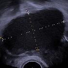
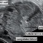
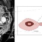
Ultrasound
Transvaginal pelvic ultrasound demonstrates an adnexal mass or cyst with a wide echogenic outer ring, either on or within the ovary . Pressure applied via the probe is unable to separate the mass from the ovary. Color Doppler may reveal a hypervascular rim (ring of fire sign). A yolk sac or embryo are uncommonly seen.
Treatment and prognosis
Like for tubal pregnancy, treatment of ovarian pregnancy is usually treated with surgical resection of the involved organ (here, oophorectomy, or wedge resection of the ovary). Medical management has been reported but realistically is reserved for cases where there is persistent trophoblastic tissue.
Differential diagnosis
In a pregnant woman without identifiable intrauterine gestational sac, an ovarian ectopic pregnancy may be misdiagnosed as the following entities that are far more common:
- corpus luteum cyst with hemorrhage or rupture
- tubal ectopic pregnancy
Siehe auch:
- tubal ectopic pregnancy
- Endometriose
- Intrauterinpessar
- Extrauteringravidität
- Unterleibsentzündung
- Eileiter
- Ovar
und weiter:


 Assoziationen und Differentialdiagnosen zu ovarian ectopic pregnancy: