intraductal papillary mucinous neoplasm (IPMN)


Intraductal papillary mucinous neoplasms or tumors (IPMNs or IMPTs) are epithelial pancreatic cystic tumors of mucin-producing cells that arise from the pancreatic ducts. They are most commonly seen in elderly patients.
On imaging, particularly MRCP, they are characterized by single or multiple unilocular or septated pancreatic cystic lesions communicating with the pancreatic ducts.
Epidemiology
These tumors are most frequently identified in older patients, 50-60 years of age , and thus are sometimes colloquially referred to as the "grandfather lesion". Main duct type (see below) appears to present a decade or so earlier on average than branch duct type . The sex distribution is roughly balanced with a possible slight male predominance .
Clinical presentation
Clinical presentation can be difficult to distinguish from chronic pancreatitis with repeated acute exacerbations. Patients can present with abdominal pain, weight loss, obstructive jaundice, pancreatitis and new-onset diabetes mellitus .
Pathology
Intraductal papillary mucinous neoplasms are one of a number of mucinous tumors of the pancreas and can be further divided both histologically and with respect to their macroscopic appearance . They are uncommon ductal epithelial tumors comprising approximately 10-15% of cystic pancreatic neoplasms.
Macroscopic appearance
Divided macroscopically:
- main duct
- reminiscent of chronic pancreatitis
- segmental or diffuse distribution
- highest malignant potential
- ~60% are malignant
- branch duct type
- mostly seen in the head and uncinate process
- more localized and mass-like
- may be multifocal
- may be macro or microcystic in appearance
- typically indolent behavior
- ~5% (range 2-10%) are malignant
- mixed-type lesions
- similar to the main duct type in terms of prognosis and overall survival
Solid components, as well as bile duct dilatation , are suspicious of malignant transformation.
Histology
They are histologically divided into:
- adenoma
- borderline-malignant
- intraductal papillary mucinous adenocarcinoma
Markers
In patients without pancreatitis, abnormal (either elevated or depressed) pancreatic enzyme markers (amylase/lipase) are associated with malignant IPMN, with the elevation of these a marker of invasiveness .
Location
Reported locations of IPMN include:
- head ~ 50%
- tail ~ 7%
- uncinate process ~ 4%,
- elsewhere throughout pancreas ~ 39%
Radiographic features
The characteristic feature is that these tumors communicate with the main pancreatic duct or its branches, which helps to distinguish these tumors from mucinous cystadenoma/cystadenocarcinoma, which do not.
ERCP
Direct imaging of the pancreatic duct demonstrates variable dilatation (segmental or diffuse or branch) depending on the type. Polypoid mural tumor or amorphous mucinous luminal filling defects may be identified .
Mucinous material may be seen protruding from the ampulla of Vater .
Ultrasound
Ultrasound may demonstrate small thin-walled pancreatic cysts or dilated hypoechoic ducts (main pancreatic duct over 5 mm in caliber). Diffuse main duct type has appearances essentially indistinguishable from chronic pancreatitis, with duct dilatation and parenchymal atrophy .
Mural nodules and mucin globules may appear hyperechoic, and difficult to separate from adjacent pancreatic parenchyma .
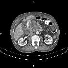
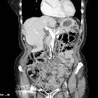
CT
They present as single or multiple pancreatic cystic hypodense lesions. Dilatation of the main duct over 5 mm is concerning for the main type IPMN. The communication with the pancreatic ducts, particularly the side branch lesions, may be difficult to demonstrate on CT. They do not calcify.
Both a dedicated pancreatic CT protocol and pancreatic MRI/MRCP have been reported having similar accuracy in the characterization of the pancreatic cystic lesions , but most recent guidelines recommend MRI as the modality of choice for IPMN followup.
MRI
MRI studies, particularly MRCP, has largely replaced CT on the imaging workup of these lesions.
- main duct IPMN (with dilatation of the main duct >5 mm)
- either segment of the pancreatic duct (or the entire duct) are dilated and filled with low density (mucin thus water signal) material
- overlying pancreatic parenchyma may be thinned
- if proximal, the distal pancreatic duct may be dilated without direct involvement (cystic neoplasms can have a similar appearance)
- solid mural nodules are concerning for malignant transformation, particularly if enhancing following administration of contrast
- occasionally mucinous material can be seen to bulge out of a dilated ampulla of Vater (uncommon but essentially pathognomonic)
- mucin globules do not enhance and lie dependently within the duct
- branch duct IPMN
- the majority of the gland is normal in appearance, except for a single or multiple side branches demonstrating marked dilatation
- cystic mass-like appearance which often mimics cystic tumors of the pancreas
- its appearance has been termed a bunch of grapes due to its appearance
- microcystic variety has appearances similar to serous cystadenomas, but again communication with the main pancreatic duct is the key to the correct diagnosis
- mixed-type IPMN
- appears like an advanced branch duct IPMN with main pancreatic duct dilatation over 5 mm
See the Tanaka criteria / International consensus guidelines for the management of IPMN and MCN of the pancreas (2012) for further details.
Treatment and prognosis
Although generally indolent, malignant degeneration does occur, with direct invasion into adjacent organs or more frequently dissemination in the peritoneal cavity (pseudomyxoma peritonei).
Current consensus criteria recommend resection for main duct IPMNs and varying treatment of branch duct IPMNs, ranging from resection to surveillance, depending on high-risk stigmata and worrisome features (see: Tanaka criteria). Patient co-morbidities and wishes clearly have a major impact on the decision to operate.
If the lesion is proximal (either segmental main duct or branch type) then a Whipple procedure may be performed. If distal then a partial pancreatectomy suffices. Complete resection is curative.
Differential diagnosis
General imaging differential considerations include:
- chronic pancreatitis
- difficult to distinguish from main duct type on account of dilated duct
- mucinous cystadenoma/cystadenocarcinoma
- should not appear to communicate with the main pancreatic duct
- serous cystadenoma
- should not appear to communicate with the main pancreatic duct
- appear similar to microcystic branch type IPMN
- 30-40% have central calcification
Practical points
The radiological report has to be clear regarding imaging worrisome features of these lesions that may guide for further surgical intervention:
- main duct IPMN
- main pancreatic duct over 5 mm
- presence of contrast-enhancing components
- branch duct IPMN
- main pancreatic duct over 5 mm
- cyst diameter ≥3 cm
- presence of a contrast-enhancing mural nodule ≥5 mm
- presence of solid mass
- thickened and enhancing cyst wall
- growth rate ≥5 mm/year
Clinical features to guide surgery may also include:


 Assoziationen und Differentialdiagnosen zu intraduktale papillär muzinöse Neoplasie:


