serous cystadenoma of pancreas


Serous cystadenoma of the pancreas, also referred as microcystic adenoma, is an uncommon type of benign cystic pancreatic neoplasm.
Epidemiology
There is a recognized strong female predilection (M:F ~ 1:4) and usually presents in middle age to elderly patients (>60 years of age).
Clinical presentation
Most patients are asymptomatic . Some may present with pain, weight loss, jaundice, or a palpable mass .
Pathology
Pancreatic serous cystadenomas are benign neoplasms composed of numerous small cysts that are arrayed in a honeycomb-like formation. There can be significant variation in locule size (1-20 mm) .
Most individual cysts are typically <10 mm .
Three morphological patterns have been described :
- polycystic: 70%
- honeycomb: 20%
- oligocystic (macrocystic variant): <10% (cysts can be larger than 20 mm)
The cysts are lined by glycogen-rich flat or cuboidal epithelium separated by fibrous septa that radiate from a central scar, which may be calcified. Lesions can be rather large at presentation (~5 cm).
Associations
- von Hippel Lindau (vHL) disease: can be multiple or diffuse and present at a younger age
Location
Lesions are distributed throughout the pancreas. In the largest series, they were found in the head/uncinate process 40% of the time, body 34%, and tail 26% .
Radiographic features
Plain radiograph
- nonspecific and will usually be normal
- may demonstrate amorphous central calcification overlying the pancreas
Ultrasound
- nonspecific hypoechoic mass in the pancreatic head region, possibly with internal echoes indicating microcysts (the oligocystic subtype may demonstrate individually identifiable cysts )
CT
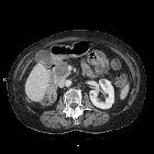
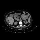
- typically demonstrates a multicystic, lobulated mass in the pancreatic head sometimes described as a 'bunch of grapes'
- the individual cysts are typically <20 mm in size and greater than six in number (except for the oligocystic variety
- a characteristic enhancing central scar may be present which can show associated stellate calcification (present in ~20% of cases)
MRI
Serous cystadenomas usually appear as a cluster of small cysts within the pancreas. There is no visible communication between the cysts and the pancreatic duct.
Signal characteristics include:
- T1: typically low signal
- T2: the central fibrous scar (if present) is of a low signal while cystic components themselves are of a high signal
- T1 C+ (Gd): fibrous septa between them may enhance on delayed contrast-enhanced images
Excluding the absence of communication with the main pancreatic duct, visualization of the lesion will not be facilitated by secretin-enhanced MRCP (SMRCP or MRCP-S) .
Angiography
- may show enhancement due to hypervascular components
Treatment and prognosis
Most lesions should be observed without treatment, unless there is diagnostic uncertainty or significant associated symptomatology . They are benign lesions and do not recur once resected .
Differential diagnosis
General imaging differential considerations on cross-sectional imaging include:
- intraductal papillary mucinous tumor (IPMN) of the pancreas: communicates with pancreatic ducts
- pancreatic pseudocyst
- mucinous cystic neoplasm of the pancreas (e.g. mucinous cystadenoma)
- calcification tends to be peripheral
- usually unilocular
- if multilocular type, individual cysts tend to be >20 mm in size
- solid pseudopapillary tumor with cystic changes and necrosis
Siehe auch:
- Pankreaspseudozyste
- zystische Pankreasläsionen
- intraduktale papillär muzinöse Neoplasie
- muzinöses Zystadenom
- muzinös zystische Neoplasien des Pankreas
- Pankreaszysten
- mikrozystisches seröres Zystadenom des Pankreas
und weiter:


 Assoziationen und Differentialdiagnosen zu seröses Zystadenom des Pankreas: