Left ventricular true aneurysm


Left ventricular aneurysms are discrete, dyskinetic areas of the left ventricular wall with a broad neck (as opposed to left ventricular pseudoaneurysms), thus often termed true aneurysms.
Epidemiology
True left ventricular aneurysms develop in less than 5% of all patients with ST-elevation myocardial infarctions (STEMI) , 5 days to three months after the infarction.
Clinical presentation
They may present with symptoms of decompensated heart failure.
ECG
- pathologic Q waves
- duration > 0.03 seconds
- depth > 1/3rd R wave amplitude
- persistent ST segment elevation
- should return to baseline within 2 weeks of a myocardial infarction
- relatively low amplitude T waves
- the ratio of precordial T wave amplitude compared to QRS amplitude should be less than 0.36
- higher T:QRS ratio implies hyperacute T waves of myocardial infarction
Pathology
The wall of the true aneurysm is thinner than the wall of the rest of the left ventricle and is usually composed of fibrous tissue as well as necrotic muscle, sometimes mixed with viable myocardium.
A true aneurysmal sac contains an endocardium, epicardium, and thinned fibrous tissue (scar) which is a remnant of the left ventricular muscle, while a pseudoaneurysm sac represents a pericardium that contains a ruptured left ventricle .
Etiology
Aneurysm formation occurs when intraventricular tension stretches the non-contracting infarcted myocardium, causing bulging of the infarcted area with each contraction.
Location
Usually, they involve the anterior or anterolateral wall.
Radiographic features
Echocardiography, cardiac MRI, and cardiac ventriculography form the mainstay of imaging. A key consideration is whether an aneurysm represents a true or false aneurysm due to the differing prognoses and management. True aneurysms typically have a wide neck.
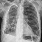
Radiography
The CXR findings of a left ventricular aneurysm are typical and include:
- focal lateral bulge arising from the left lower heart border
- may have thin curvilinear calcification in the wall of the aneurysm
- left retrocardiac double density (particularly pseudoaneurysms)
Echocardiography
The ratio of the maximum diameter of the orifice to the maximum internal diameter of the cavity may higher (0.9-1.0) than for a pseudoaneurysm .
Treatment and prognosis
The rate of mortality in patients with left ventricular aneurysms is up to 6x higher than in patients without aneurysms. Death is often sudden and may be related to the high incidence of ventricular tachyarrhythmias associated with these aneurysms .
Complications
Recognized complications include:
- intramural thrombus, which may calcify
- impaired cardiac output
- aneurysm rupture: rare
- rupture rate is higher with pseudoaneurysms
Differential diagnosis
In certain situations consider:
Siehe auch:
- Herzspitzenaneurysma
- Verkalkungen im Röntgenbild des Thorax
- Herzwandaneurysma
- Pseudoaneurysma des linken Ventrikels
und weiter:


 Assoziationen und Differentialdiagnosen zu Aneurysma des linken Ventrikels: