maligner rhabdoider Tumor der Niere


Rhabdoid tumor of the kidney is a rare, highly aggressive malignancy of early childhood, closely related to atypical teratoid/rhabdoid tumors (AT/RT) of the brain (see rhabdoid tumors).
Epidemiology
Rhabdoid tumors occur exclusively in children, with 60% occurring before the age of 1 year of age, and 80% before the age of 2 years (average age of presentation = 11 months). Approximately 2% of pediatric renal malignancies are rhabdoid tumors .
Clinical presentation
Rhabdoid tumors are very aggressive and initial manifestation may be due to metastatic disease. Local symptoms and signs include hematuria and loin/flank mass. Additionally, patients may develop hypercalcemia secondary to elevated parathormone levels .
Pathology
The term "rhabdoid" stems from the histologic appearance, which resembles that of a tumor of skeletal muscle origin, although in fact, rhabdoid cells are a distinct cellular population. All rhabdoid tumors share deletions in the long arm of chromosome 22, mapped to the INI-1 gene, believed to be a tumor suppressor .
Radiographic features
On all modalities, rhabdoid tumors appear as large, centrally located, heterogeneous soft-tissue masses, involving the renal hilum with indistinct margins.
CT
Rhabdoid tumors are large and heterogeneous, usually located centrally within the kidney. They are lobulated with individual lobules separated by intervening areas of decreased attenuation, relating to either previous hemorrhage or necrosis . Enhancement is similarly heterogeneous.
Calcification is relatively common, seen in up to 66% of cases and is typically linear and tends to outline tumor lobules .
Subcapsular fluid accumulation is said to be a relatively characteristic feature, not often seen in other pediatric renal tumors. Although, due to the rarity of renal rhabdoid tumors, a pediatric renal neoplasm with a subcapsular fluid collection is still more likely to represent Wilms tumor .
Treatment and prognosis
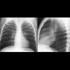
Rhabdoid tumors have the worst prognosis of all renal tumors. It is highly aggressive and metastasizes early, with up to 80% of patients presenting with metastatic disease, typically to the lungs and less often to the liver, abdomen, brain, lymph nodes, or skeleton .
Treatment consists of radical nephrectomy and resection of adjacent lymph nodes followed by chemotherapy. Survival is poor, with an 18-month survival rate of only 20%, and almost all patients succumbing before 5 years post-diagnosis .
Differential diagnosis
The main differential is that of the far more common Wilms tumor. Although imaging appearances can be similar, a number of features can help point towards the diagnosis of a rhabdoid tumor. These include :
- subcapsular fluid collections
- tumor lobules separated by hypoattenuating areas of necrosis or hemorrhage
- calcifications
- more common than in Wilms tumors (66% in rhabdoid vs 6-15% in Wilms)
- typically linear distribution outlining tumor lobules
- vascular and local invasion is more common
- a synchronous intracranial neoplasm is a distinguished feature of rhabdoid tumor
Siehe auch:
- Nephroblastom
- pädiatrische Nierentumoren
- kongenitales mesoblastisches Nephrom
- maligner rhabdoider Tumor
und weiter:

 Assoziationen und Differentialdiagnosen zu maligner rhabdoider Tumor der Niere:


