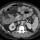
Mesenterialvenenthrombose


Veno-occlusive mesenteric ischemia is most often the result of superior mesenteric vein (SMV) thrombosis and is a less common cause of acute mesenteric ischemia. Often despite thrombosis of the SMV, small bowel necrosis does not occur, presumably due to persistent arterial supply and some venous drainage via collaterals.
Epidemiology
Compared to acute superior mesenteric artery occlusion, veno-occlusive causes of acute mesenteric ischemia are uncommon, accounting for only 5-15% of all cases of acute mesenteric ischemia .
Clinical presentation
Acute superior mesenteric vein thrombosis presents vaguely as an acute abdomen with gradually worsening diffuse, colicky abdominal pain, associated with distention, and symptoms may have been present for a few days . Heme-positive stool may also be present . As ischemia progresses, eventual necrosis, perforation, sepsis and shock ensue.
Pathology
As the superior mesenteric vein thrombosis, back pressure builds as arterial supply to the bowel is uninterrupted. This leads to bowel wall edema (and thus bowel wall thickening) and possible intramural hemorrhage (leading to hyperdense bowel wall) . Depending on how complete the occlusion is, and whether or not collateral drainage is available, the combination of backpressure, and bowel wall thickening can lead to inadequate tissue perfusion leading to intestinal ischemia, infarction and eventual necrosis .
Etiology
The majority of cases are considered secondary to an identifiable underlying condition, including :
- hypercoagulable states
- recent abdominal surgery
- intra-abdominal or systemic sepsis
- portal hypertension
- mechanical narrowing due to adjacent malignancy
Approximately 30% (range 20-40%) of cases have no clear precipitant and are considered idiopathic .
Radiographic features
Imaging is the only reliable way of making the diagnosis, especially as clinical presentation is vague. CT is the most accurate test available to us at present, with excellent sensitivity (up to 100%). Ultrasound and catheter mesenteric angiography are reported to have ~70% sensitivity but in practice are infrequently requested as a first-line investigation, unless the presentation suggests another diagnosis (e.g. cholecystitis).
CT
Technique
For a discussion on CT technique refer to intestinal ischemia article.
Findings
The findings in superior mesenteric vein thrombosis include :
- vascular changes
- filling defect in the superior mesenteric vein and branches (seen in 90% of cases)
- mesenteric congestion and stranding
- bowel changes
- typically thickened (most common finding): due to hemorrhage or edema
- 8-9 mm (usually <15 mm)
- normal wall thickness is 3-4 mm
- target sign
- density (variable)
- hypoattenuating due to edema
- hyperdense wall on non-contrast images (due to intramural hemorrhage)
- enhancement (variable)
- absent once infarcted (more common in arterial occlusion)
- hyperenhancement or target appearance
- prolonged enhancement
- pneumatosis intestinalis: due to transmural infarction
- dilated and fluid-filled lumen
- typically thickened (most common finding): due to hemorrhage or edema
- other changes
- ascites
Treatment and prognosis
Traditionally management has been surgical, with assessment of the small bowel for necrosis and resection of necrotic bowel, followed by anticoagulation. This has a reported mortality rate of 7-20% , which, although still high, is much better than 92-100% mortality with conservative management .
Surgery is clearly still required in patients with intestinal infarction; however, in patients where this has not yet occurred, endovascular thrombolysis/thrombectomy is a viable option . Thrombolytic agents can be introduced either via a superior mesenteric artery catheter or a transhepatic transportal retrograde approach, whereas mechanic clot lysis can only be achieved via a retrograde transhepatic venous approach .
Probably the safest way to directly access the portal circulation is via a transjugular-transhepatic approach (similar to the initial steps of a TIPS). A direct transhepatic approach has also been performed but is associated with a greater risk of hemorrhage .
Complications
Complications include :
- surgical complications
- abscess
- anastomosis breakdown
- short bowel syndrome
- recurrent mesenteric thrombosis: 14%, usually within six weeks
Differential diagnosis
If a superior mesenteric vein thrombus/filling defect can be identified, no differential diagnosis exists. In cases where a filling defect cannot be identified, the differential diagnosis is essentially that of bowel wall thickening and includes:
- vascular
- acute superior mesenteric artery occlusion
- ischemia due to hypotension
- submucosal hemorrhage or hematoma
- inflammation/infection
- neoplasm
- pseudothickening related to incomplete distention and residual fluid
Siehe auch:
- Morbus Crohn
- Mesenterialinfarkt
- Pfortaderthrombose
- Dünndarmischämie
- neutropene Enterokolitis
- radiation enteritis
- akuter Verschluss der Arteria mesenterica superior
und weiter:
- Vena mesenterica superior
- Gewebsreaktion Fett Bauchraum
- porto-mesenteric venous thrombosis in Crohn's disease
- Mesenterialvenenenge
- intestinal venous infarction
- Mesenterialverschluss
- Dünndarmischämie bei Mesenterialvenenthrombose
- porto-mesenteric venous thrombosis
- Mesenterialvarikosis
- superior mesenteric vein thrombosis after a chemotherapy

 Assoziationen und Differentialdiagnosen zu Mesenterialvenenthrombose:


