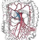
Arterial occlusive mesenteric ischemia
Arterial occlusive mesenteric ischemia can be a life-threatening event related to obstruction of the mesenteric arteries, most commonly the superior mesenteric artery (SMA), supplying the small bowel and colon. It is the most common cause of mesenteric ischemia.
Epidemiology
An acute occlusion is an uncommon event that typically affects elderly patients, who are at an increased risk of other cardiovascular events.
Risk factors
- advanced age
- smoking
- prothrombotic tendency
- antiphospholipid antibodies, etc.
- valvular/cardiac abnormalities
- mechanical heart valve
- atrial fibrillation
- acute myocardial ischemia
- ventricular aneurysm
- right-to-left shunt
- patent foramen ovale/atrial septal defect with paradoxical embolism
Clinical presentation
Clinical presentation is variable and unfortunately often non-specific such that the diagnosis is not made for some time. It may be dramatic with acute onset severe abdominal pain or can be less well-defined .
Pathology
Acute arterial occlusion can be due to a number of causes :
- embolic event: ~50% (range 40-60%)
- acute in situ thrombosis superimposed on atherosclerosis: 15-30%
- aortic dissection with the involvement of the SMA origin
- slow flow or idiopathic
Radiographic features
Ultrasound
Ultrasound is able to demonstrate normal flow in both the SMA and SMV but is incapable of assessing side branches or the bowel wall. It has little role in the acute management of this condition.
CT
Computed tomography is widely accepted as the first-line imaging technique for evaluation due to its speed, widespread availability and ability to diagnose alternative causes of acute abdominal pain.
Technique
For a discussion on CT technique, refer to the mesenteric ischemia article.
Findings
Findings in acute SMA occlusion include :
- arterial changes: lack of enhancement of the lumen of the SMA and/or its branches
- embolism location varies
- 15% at the origin of the SMA
- 50% immediately distal to the origin of the middle colic artery
- thrombosis usually occurs in the proximal 2 cm of the SMA
- embolism location varies
- bowel changes: reflecting reduction/obliteration of blood supply
- mucosal/serosal enhancement absent
- thickness
- variable
- in pure arterial occlusion, the wall may be thinned (a.k.a. paper-thin wall) due to loss of intestinal muscular tone and absence of blood supply
- a thickened wall may also be present but does not correlate with severity; reperfusion can cause a thickened wall
- ileus / dilated loops of bowel: >3 cm in diameter
- air-fluid or blood-fluid levels: due to dysfunctional peristalsis
- necrotic mural gas may be present: pneumatosis intestinalis
- other changes
- mesenteric edema
- free fluid
- intrahepatic portal venous gas: due to pneumatosis intestinalis
- free intra-abdominal gas
- causes: e.g. intracardiac thrombus in a dilated left ventricle
Angiography (DSA)
Once the gold standard for diagnosis, now reserved for patients who may benefit from endovascular intervention.
Treatment and prognosis
An acute SMA occlusion carries a mortality of 75-90% despite treatment . Treatment options include :
- endovascular thrombectomy
- intraluminal papaverine
- surgical thrombectomy with resection of non-salvageable infarcted bowel
Differential diagnosis
- mesenteric arteritis
- splanchnic venous occlusion: superior mesenteric vein or portal vein thrombosis rather than arterial occlusion
- chronic arterial occlusion with an alternative cause for abdominal pain: identification of well-formed collaterals may suggest that the occlusion is chronic
- small bowel obstruction
- Crohn disease: in most cases, a significantly different patient group
Siehe auch:
- Pneumatosis intestinalis
- Morbus Crohn
- Dünndarmileus
- Mesenterialinfarkt
- Arteria-mesenterica-superior-Syndrom
- Pfortaderthrombose
- Atriumseptumdefekt
- Kolon
- Arteria mesenterica superior
- Dünndarm
- chronische mesenteriale Ischämie
- Verschluss der Arteria mesenterica superior
und weiter:

 Assoziationen und Differentialdiagnosen zu akuter Verschluss der Arteria mesenterica superior:


