mesenteric neoplasms ct appearances of primary and secondary tumors and differential diagnosis
Solitärer
fibröser Tumor des Omentum in der Computertomografie oben axial arteriell und portalvenös, unten koronar und sagittal portalvenös. Beachte auch den Gefäßstiel.

Sandwich sign
(mesentery) • Lymphoma - sandwich sign - Ganzer Fall bei Radiopaedia

A case of
mesenteric teratoma. Non-enhanced CT: evidence of foci of calcification within the mass.

A case of
mesenteric teratoma. Coronal reformation: the small bowel loops were displaced to the left side

This image
is part of a series which can be scrolled interactively with the mousewheel or mouse dragging. This is done by using Template:Imagestack. The series is found in the category Peritoneal mesothelioma - CT - case 001. Peritoneales desmoplastisches Mesotheliom in der Computertomographie. Dieses Bild ist Teil einer Serie zum Durchblättern (siehe Kategorie).

Malignant
peritoneal mesothelioma • Primary peritoneal mesothelioma - Ganzer Fall bei Radiopaedia

Detection of
peritoneal metastases. Lymphatic metastases. 45-year-old male patient with Non-Hodgkins Lymphoma showing the characteristic ‘Sandwich’ sign of mesenteric and retroperitoneal lymph node involvement.

A case of
mesenteric teratoma. Post contrast image showing intraperitoneal mass containing solid, cystic and fatty elements and foci of calcifications.

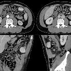
A giant
solitary fibrous tumor of the mesentery: a case report and literature review. (A) Abdominal computed tomography demonstrated a 25 × 11 cm, heterogeneous, lobulated mass in the abdominal cavity. (B) Colonal view demonstrated lobulated mass.

Mesenteric
cystic lymphangioma. CT with i.v. contrast shows a mesenteric cystic mass - density of the fluid within the lesion was 8-10 HU.

Mesenteric
cystic lymphangioma in adult: a case series and review of the literature. MRI of the abdomen. Axial (a) and coronal (b) T2-weighted TSE images showed the presence of a fluid-filled cystic lesion with a diameter of 35 mm in the retrocecal adipose tissue.

Mesenteric
cystic lymphangioma in adult: a case series and review of the literature. CT of the abdomen. Axial (a) and MPR coronal (b) CT images showed a huge mass composed by multiple confluent cystic lesions occupying the peritoneal spaces and displacing inferiorly the small bowel.
Neoplasien des Mesenteriums
mesenteric neoplasms ct appearances of primary and secondary tumors and differential diagnosis
Siehe auch:
- mesenteriale Pannikulitis
- peritoneales Mesotheliom
- mesenteriales Lymphom
- zystisches Lymphangiom des Mesenterium
- mesenteriales Teratom
- mesenteric and retroperitoneal lymphoma
- Liposarkom des Mesenteriums
- abdominelles Liposarkom
- mesenteriales Hibernom
- mesenteriales Sarkom
- malignant solitary fibrous tumour of the mesentery
- Lymphangiomyom des Mesenteriums
- Desmoid-Tumor des Mesenteriums
- Karzinoid des Gastrointestinaltraktes
- mesenterialer gastrointestinaler Stromatumor
- solitärer fibröser Tumor des Peritoneums
und weiter:


 Assoziationen und Differentialdiagnosen zu Neoplasien des Mesenteriums:


