Neoplasien des Mesenteriums
Solitärer
fibröser Tumor des Omentum in der Computertomografie oben axial arteriell und portalvenös, unten koronar und sagittal portalvenös. Beachte auch den Gefäßstiel.

Sandwich sign
(mesentery) • Lymphoma - sandwich sign - Ganzer Fall bei Radiopaedia

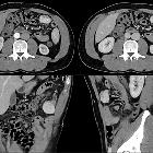
A case of
mesenteric teratoma. Non-enhanced CT: evidence of foci of calcification within the mass.

A case of
mesenteric teratoma. Coronal reformation: the small bowel loops were displaced to the left side

This image
is part of a series which can be scrolled interactively with the mousewheel or mouse dragging. This is done by using Template:Imagestack. The series is found in the category Peritoneal mesothelioma - CT - case 001. Peritoneales desmoplastisches Mesotheliom in der Computertomographie. Dieses Bild ist Teil einer Serie zum Durchblättern (siehe Kategorie).

Malignant
peritoneal mesothelioma • Primary peritoneal mesothelioma - Ganzer Fall bei Radiopaedia

Detection of
peritoneal metastases. Lymphatic metastases. 45-year-old male patient with Non-Hodgkins Lymphoma showing the characteristic ‘Sandwich’ sign of mesenteric and retroperitoneal lymph node involvement.

A case of
mesenteric teratoma. Post contrast image showing intraperitoneal mass containing solid, cystic and fatty elements and foci of calcifications.

A giant
solitary fibrous tumor of the mesentery: a case report and literature review. (A) Abdominal computed tomography demonstrated a 25 × 11 cm, heterogeneous, lobulated mass in the abdominal cavity. (B) Colonal view demonstrated lobulated mass.

Mesenteric
cystic lymphangioma. CT with i.v. contrast shows a mesenteric cystic mass - density of the fluid within the lesion was 8-10 HU.

Mesenteric
cystic lymphangioma in adult: a case series and review of the literature. MRI of the abdomen. Axial (a) and coronal (b) T2-weighted TSE images showed the presence of a fluid-filled cystic lesion with a diameter of 35 mm in the retrocecal adipose tissue.

Mesenteric
cystic lymphangioma in adult: a case series and review of the literature. CT of the abdomen. Axial (a) and MPR coronal (b) CT images showed a huge mass composed by multiple confluent cystic lesions occupying the peritoneal spaces and displacing inferiorly the small bowel.
Neoplasien des Mesenteriums
Siehe auch:
- mesenteriale Pannikulitis
- peritoneales Mesotheliom
- zystisches Lymphangiom des Mesenterium
- mesenteriales Lymphom
- mesenteriales Teratom
- mesenteric and retroperitoneal lymphoma
- Liposarkom des Mesenteriums
- mesenteriales Sarkom
- abdominelles Liposarkom
- mesenteriales Hibernom
- malignant solitary fibrous tumour of the mesentery
- Desmoid-Tumor des Mesenteriums
- Lymphangiomyom des Mesenteriums
- Karzinoid des Gastrointestinaltraktes
- mesenterialer gastrointestinaler Stromatumor
- solitärer fibröser Tumor des Peritoneums
und weiter:


 Assoziationen und Differentialdiagnosen zu Neoplasien des Mesenteriums:


