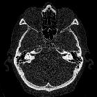
Michel deformity

Michel aplasia or deformity, also known as complete labyrinthine aplasia, is the most severe congenital inner ear malformation, characterized by complete absence of inner ear structures (cochlea, vestibule, semicircular canals, and vestibular and cochlear aqueducts).
Epidemiology
It is extremely rare, accounting for less than 1% of inner ear malformations .
Associations
- abnormal development of the skeletal portions of the second arch
- non-differentiation of the stapes, with resultant absence of round and oval windows
- abnormal course of the facial nerve
- skull base abnormalities
- hypoplasia of the petrous temporal bone; hypoplastic and sclerotic petrous apex may mimic labyrinthitis ossificans
- platybasia
- aberrant course of the jugular vein
Pathology
Michel aplasia is thought to result from failure of development of the otic placode at or before the 3 week of gestation .
Classification
Sennaroglu described three subgroups based on radiological findings :
- with hypoplastic or aplastic petrous bone: the petrous part of the temporal bone is small or absent and the middle ear is closer to the posterior fossa
- without otic capsule: the petrous bone is normal in size but the otic capsule within is small or absent
- with otic capsule: the petrous bone and otic capsule are normally formed, even as the internal contents have not developed; the labrythine segment of the facial nerve canal may be normal in position
Radiographic features
CT
The finding is typically bilateral . In unilateral cases, the other side typically has another form of severe dysplasia .
The internal auditory canal is absent or atretic .
There is no lucency in the location of the otic capsule in the petrous temporal bone to indicate labyrinthine structures.
MRI
The vestibulocochlear nerve is not detectable .
History and etymology
P Michel first described the condition in 1863 .
Siehe auch:
und weiter:

 Assoziationen und Differentialdiagnosen zu Michel-Aplasie:


