nutcracker syndrome


 nicht verwechseln mit: Nussknackerösophagus
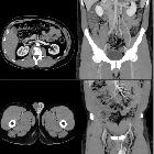
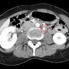
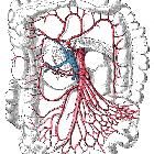
Nutcracker syndrome is a vascular compression disorder that refers to the compression of the left renal vein most commonly between the superior mesenteric artery (SMA) and aorta, although other variations can exist . This can lead to renal venous hypertension, resulting in rupture of thin-walled veins into the collecting system with resultant hematuria.
Terminology
In certain situations, the syndrome can result from a retroaortic or circumaortic left renal vein.
Nutcracker syndrome should not be confused with superior mesenteric artery syndrome (Wilkie syndrome) also a superior mesenteric artery compression disorder, where the SMA compresses the third part of the duodenum (the two conditions, however, may be associated).
Epidemiology
There is a slightly greater female predilection.
Clinical presentation
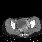
The most common clinical manifestations of nutcracker syndrome are left flank pain, pelvic pain, hematuria and gonadal varices. Orthostatic proteinuria has also been reported. Hematuria can be microscopic or macroscopic. Hematuria should be from the left ureteric orifice only . In the absence of clinical symptoms, renal vein compression is referred to as nutcracker phenomenon or nutcracker anatomy, which can be a more common situation.
Pathology
Associations
- may occur simultaneously with SMA syndrome
- an association with a thin or asthenic body habitus has long been noted.
Types
Compression of the left renal vein can occur primarily in two anatomic locations .
- anterior nutcracker syndrome (classical): occurs at the branching of the SMA off of the aorta
- posterior nutcracker syndrome (rarer form): a retroaortic left renal vein is compressed between the aorta and vertebrae
- rarely, a combination form may occur
Radiographic features
Radiographic features are similar on ultrasound, Doppler ultrasound, CT, MRI, and conventional angiography:
For classical form
- reduced aortic-SMA angle: normal angle between aorta and SMA is ~45° (range 38-65°)
- left renal vein stenosis
- collateral pathways: main collateral pathway is the left gonadal vein which will display early enhancement during portal venous phase
- pressure gradient >3 mmHg on renal venography in early-stage nutcracker syndrome; well-developed collateral veins dissipate the high-pressure gradient
- compression ratio (CR) given by the anteroposterior diameter of the precompressed vein (P) divided by that of the compressed vein (C); namely, CR = P/C
- a compression ratio above 2.25 is highly sensitive and specific for nutcracker syndrome
Complications
Persistent hematuria can precipitate renal vein thrombosis .
Treatment and prognosis
Treatment should be started strictly when it is causing symptoms (hematuria and left flank pain). Surgical treatment modalities have their inherent complications and should be contemplated only when strongly indicated. A few of the reported surgical choices are:
- left renal vein transposition
- kidney autotransplant
- superior mesenteric artery transposition
- gonadal vein transposition
- nephrectomy
- extravascular stent placement
- endovascular stent-graft placement
History and etymology
The first clinical report of this syndrome was made by El-Sadr and Mina in 1950 while the term "nutcracker syndrome" is thought to have been first used by de Schepper in 1972.
See also
Siehe auch:
- Aorta
- Arteria-mesenterica-superior-Syndrom
- Nierenvenenthrombose
- Arteria mesenterica superior
- Ovarialvenenthrombose
- Varicocele der Vena ovarica
- vaskuläre Kompressionssyndrome
- circumaortic renal collar
- superior mesenteric artery compression disorders
- spontane Ruptur der Vena ovarica
- Varizen der Labien
und weiter:

 Assoziationen und Differentialdiagnosen zu Nussknacker-Syndrom:


