Ösophagitis

Esophageal
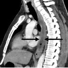
emergencies: another important cause of acute chest pain. Acute esophagitis. Fifty-year-old male with diffuse chest pain and mild fever. Sagittal CT image show diffuse circumferential wall thickening with mild enhancement involving almost entire esophagus (arrows). Intraluminal fluid (asterisk) is noted

Esophagitis
• Esophagitis - Ganzer Fall bei Radiopaedia

Candida-Ösophagitis
in der Röntgenbreischluckuntersuchung. Induration mit Verminderung / Aufhebung der Peristaltik und Nischenbildung in den Ulcera.
Esophagitis refers to inflammation of the esophagus. It can arise from a range of causes which include:
- infective esophagitis
- non-infective esophagitis
Radiographic features
Fluoroscopy
Radiographic signs of esophagitis depend on the fluoroscopic technique used, but include :
- mucosal irregularity
- erosions and ulcerations
- abnormal motility
- thickened esophageal folds (>3 mm)
- limited esophageal distensibility
- esophageal strictures
- intramural pseudodiverticulosis
- ulcers are a hallmark finding of esophagitis
- small ulcers (<1 cm)
- reflux esophagitis
- Herpes esophagitis
- acute radiation-induced esophagitis
- drug-induced esophagitis
- benign mucus membrane pemphigoid
- larger ulcers (>1 cm)
- small ulcers (<1 cm)
Siehe auch:
- Intramurale Pseudodivertikulose des Ösophagus
- gastroösophageale Refluxerkrankheit
- candida oesophagitis
- eosinophile Ösophagitis
- postaktinische Ösophagitis
- kaustische Ösophagitis
- Morbus Crohn des Ösophagus
- virale Ösophagitis
und weiter:

 Assoziationen und Differentialdiagnosen zu Ösophagitis:


