Osteoid Osteoma Radio-frequency Ablation

Osteoid
osteoma of the femoral head treated by radiofrequency ablation: a case report. Plain radiograph in anteroposterior view showing a well defined lytic lesion with a thin sclerotic rim located in the subarticular portion of the left femoral head (white arrow).

Osteoid
osteoma of the femoral head treated by radiofrequency ablation: a case report. T1-weighted axial MRI showing a corresponding hypointense lesion (white arrow).

Osteoid
osteoma of the femoral head treated by radiofrequency ablation: a case report. T2-weighted coronal image showing hyperintense focus with a hypointense rim (black arrows).

Osteoid
osteoma of the femoral head treated by radiofrequency ablation: a case report. T2 fat-suppressed images in coronal sections showing hyperintense focus with a hypointense rim (black arrows).

Osteoid
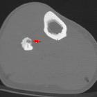
osteoma of the femoral head treated by radiofrequency ablation: a case report. Axial computed tomography section confirming a clearly defined lucent nidus with surrounding sclerotic rim (white arrow).

Osteoid
osteoma of the femoral head treated by radiofrequency ablation: a case report. Radionuclide bone scan demonstrating corresponding focal hot spot (black arrow).

Osteoid
osteoma of the femoral head treated by radiofrequency ablation: a case report. Axial computed tomography section with radiofrequency ablation (RFA) needle placed within the drilled tract.

Osteoid
osteoma of the femoral head treated by radiofrequency ablation: a case report. Residual air pockets post radiofrequency ablation.
Osteoid Osteoma Radio-frequency Ablation
Radiofrequenzablation (RFA) Radiopaedia • CC-by-nc-sa 3.0 • de
Radiofrequency ablation (RFA) is a medical procedure that uses a high-frequency alternating current produced by a radiofrequency generator oscillating in a closed-loop circuit. This current heats a needle to over 60°C, which is used to cause intentional protein denaturation and tissue damage .
Applications
- tumor ablation: established for the local destruction of liver, lung, and bone tumors, e.g. HCC and osteoid osteoma
- cardiac arrhythmia: used in the treatment of an abnormally discharging focus within the heart
- ablation neurotomy and pain management for specific nerve plexuses or Morton neuroma
- varicose veins: an alternative to sclerotherapy, as the produced heat causes closure of the affected vein
Complications
- thermal injuries to the adjacent structures: in liver tumor ablation, this means potential injury to the diaphragm, colon, gallbladder, or bile ducts, which may result in perforation, leakage of secretions, or stricture formation
- pneumothorax and pleural effusion: in lung tumor ablation
- hemorrhage: small vessels are more liable to hemorrhage more than large vessels which are rather protected by "heat sink" effect of flowing blood
- infection is a relatively low risk of radiofrequency ablation
- tumor seeding along the needle tract
- post-ablation syndrome: flu-like symptoms, including low-grade fever, chills, nausea, pain, and malaise. It is a self-limited condition that usually begins three days after ablation
- dysfunction of a cardiac pacemaker or defibrillator due to the use of radiofrequency waves
Siehe auch:

 Assoziationen und Differentialdiagnosen zu Osteoid Osteoma Radio-frequency Ablation:

