paediatric renal tumours and masses

Newborn with
a palpable right sided abdominal massTransverse (above) and sagittal (below) US images shows an enlarged solid reniform shaped mass in the right renal fossa.The diagnosis was mesoblastic nephroma.

Mesoblastic
nephroma • Mesoblastic nephroma - Ganzer Fall bei Radiopaedia

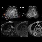
Toddler with
presumed urinary tract infection. Sagittal US of the left kidney (upper left) shows a round hyperechoic lesion in the lower pole of the kidney. Axial T2 MRI without contrast of the abdomen (upper right) and coronal T1 MRI without (lower left) and with (lower right) contrast of the abdomen shows a well-circumscribed, solid T1 hypointense and T2 isointense mass in the lower pole of the left kidney that enhances minimally.The diagnosis was Wilms tumor of the left kidney.

Toddler with
a left abdominal mass. The diagnosis was Wilms tumor of the left kidney and nephrogenic rests from nephroblastomatosis in the right kidney.The diagnosis was Wilms tumor of the left kidney and nephrogenic rests from nephroblastomatosis in the right kidney.

Wilms tumoru
with bony-mandibular, rib and calvarial metastases. A large heterogeneously enhancing mass seen in upper pole and interpolar region of right kidney. A lytic lesion in the right second rib with adjacent large soft tissue is also seen.

Synchronous
choroid plexus papilloma and Wilms tumor in a girl, disclosing a Li-Fraumeni syndrome. Radiological studies. Panel a Intravenous contrast-enhanced abdominal CT, coronal view: a solid, heterogeneous tumor in the upper pole of the left kidney is identified (upper, thin arrow), with an adjacent subcapsular perirenal hematoma (lower, thick arrow). The actual diameters were 36 mm × 28 mm × 27 mm in anteroposterior, transverse, and craniocaudal planes. Panel b Cerebral MRI, T2-weighted sequence, axial view. A solid supratentorial intraventricular tumor with left parietal lobe extension is shown. Cystic areas in the basal peripheral and medial aspects are identified .The tumor causes localized ventriculomegaly, vasogenic edema and midline shift. The actual diameters (tumor and cysts) were 81 × 59 × 75 mm in the anteroposterior, transverse, and craniocaudal planes. Panel c Non-enhanced Brain CT scan after shunt insertion. Axial reconstruction. A left parietal tumor with extensive calcification is shown. Ventricular catheter tip is in the right frontal ventricular horn

Wilms tumor
• Wilms tumor - Ganzer Fall bei Radiopaedia

Wilms tumor
• Wilms tumor - Ganzer Fall bei Radiopaedia

Wilms tumor
• Wilms tumor - Ganzer Fall bei Radiopaedia

Wilms tumor
• Wilms tumor - Ganzer Fall bei Radiopaedia

Wilms tumor
• Wilms tumor - Ganzer Fall bei Radiopaedia

Nephroblastomatosis
• Nephroblastomatosis - Ganzer Fall bei Radiopaedia

Nephroblastomatosis
• Nephroblastoma - bilateral - Ganzer Fall bei Radiopaedia

Congenital
tumors: imaging when life just begins. Renal tumors. Case 1. Mesoblastic nephroma. Axial CECT (a) and fat-saturated T1-weighted image after i.v. contrast medium administration (b) show a solid, focal renal mass replacing the normal right renal parenchyma (block arrows) in this newborn boy. Case 2. Nephroblastomatosis with bilateral Wilms’ tumors. Axial CECT (c) shows the well-defined bilateral renal masses (stars) in this 3-month-old baby, corresponding to a histologically proven bilateral Wilms’ tumor in a child with areas of nephroblastomatosis. The masses remain hypodense compared with normal renal parenchyma. Wilms’ tumors are very rare in fetuses and neonates. Case 3. MCN. T2-HASTE MR image (d) shows a multilocular cystic mass in the right kidney with tumor septae. Observe a similar but smaller left renal lesion (white arrow) in this 10-week-old boy with multiple congenital tumors (same patient as in Fig. 6c)

Rhabdoid
renal tumor: an aggressive embryonal tumor in an infant — a case report. Ultrasound picture: revealing a 7 cm heterogeneous spherical mass arising from the apical pole of the right kidney with multiple hemorrhagic areas and fine calcifications

Rhabdoid
renal tumor: an aggressive embryonal tumor in an infant — a case report. A CT scan: revealing an important uretero-pyelo-calicielle dilation with fine calcifications
pädiatrische Nierentumoren
paediatric renal tumours and masses
Siehe auch:
- Angiomyolipom der Niere
- Nierenzellkarzinom
- Neuroblastom
- Nephroblastomatose
- Nephroblastom
- medulläres Nierenkarzinom
- multilokuläres zystisches Nephrom
- Neuroblastom vs Nephroblastom
- kongenitales mesoblastisches Nephrom
- multicystic dysplastic kidneys
- maligner rhabdoider Tumor der Niere
und weiter:

 Assoziationen und Differentialdiagnosen zu pädiatrische Nierentumoren:


