Papillary tumor of the pineal region


Papillary tumors of the pineal region (PTPR) are a relatively recently described entity, and considered one of four pineal parenchymal tumors under the current (2016) WHO classification of CNS tumors.
Epidemiology
Papillary tumor of the pineal region are seen in a wide range of ages, reported from 1-70 years of age, with most cases found in middle age . They are encountered equally in both genders .
Clinical presentation
As with all other pineal region masses clinical presentation is mainly from obstructive hydrocephalus secondary to compression of the tectum of the midbrain and obstruction of the aqueduct. Compression of the superior colliculi can also lead to a characteristic gaze palsy, known as Parinaud syndrome.
Pathology
Papillary tumor of the pineal region are though to arise from specialized ependymocytes of the subcommissural organ located in the lining of the posterior commissure rather than from the pineal gland itself . It is also hypothesized that some, or perhaps many, previously published descriptions of unusual pineal region tumors (e.g. choroid plexus papillomas and papillary ependymomas) actually represent PTPRs, as histologically they are difficult to distinguish, requiring immunohistochemistry .
These tumors are considered WHO grade II or III tumors .
Macroscopic appearance
Papillary tumor of the pineal region are macroscopically indistinguishable from pineocytomas, appearing as well circumscribed tumors of the pineal region .
Microscopic appearance
Papillary tumor of the pineal region demonstrate varible morphology ranging from solid to predominantly papillary, reminiscent of ependymomas, including the presence of ependymal rosettes. Areas of necrosis are sometimes identified .
Immunophenotype
PTPRs have a fairly characteristic immunohistochemical profile that allows them to be distinguished from other pineal parenchymal tumors.
- cytokeratins (AE1/3, CAM5.2, KL1, CK18): positive
- S100: positive
- vimentin: positive
- tansthyretin: positive
- neuron-specific enolase: positive
- MAP2: positive
- GFAP: variable
Radiographic features
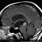
MRI
Papillary tumors of the pineal region are often indistinguishable from pineocytomas. Sometimes they demonstrate intrinsic high T1 signal attributed to secretory inclusions . This is a relatively specific findings when other causes of T1 shortening are excluded (e.g fat in a teratoma or lipoma, melanoma, hemorrhagic metastases) .
Screening of the entire neural axis is required as CSF dissemination has been reported in up to 7% of cases .
Differential diagnosis
On imaging consider other pineal region masses, particularly:
- pineocytoma and pineal parenchymal tumor of intermediate differentiation: indistinguishable from each other based only on imaging and immunohistochemistry tests are required
Siehe auch:
- Tumoren der Pinealisregion
- Pineoblastom
- Pineozytom
- Verschlusshydrocephalus
- intrakranielles Germinom
- Aquäduktsyndrom
- WHO classification
und weiter:


 Assoziationen und Differentialdiagnosen zu Papillärer Tumor der Pinealisregion: