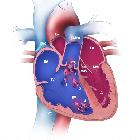
quadricuspid aortic valve
Quadricuspid aortic valve (QAV) is a rare cardiac valvular anomaly where the aortic valve has four cusps, instead of the usual three.
Epidemiology
The estimated incidence on necropsy at ~1 in 8,000. While the incidence of QAV on 2D echocardiography has been reported to range between 0.01-0.04%. There is no recognized gender predilection. Some reports suggest that this anomaly may be in up to 1% of individuals who present for aortic valve surgery .
Associations
A quadricuspid aortic valve is usually isolated but can be occasionally associated with other cardiac anomalies, such as:
- congenital coronary artery anomalies (e.g. single orifice for the coronary arteries or presence of an accessory artery) and displacement of the coronary ostia because of the accessory cusp
- hypertrophic cardiomyopathy
- subaortic stenosis
- patent ductus arteriosus
- ventricular septal defects
- ruptured sinus of Valsalva
- complete heart block
- endocarditis
Pathology
Sub types
Several variations of QAV are described. The method that was traditionally described by Hurwitz and Roberts includes :
- type a: four equal-sized cusps
- type b: three equal-sized cusps and one smaller cusp, considered commonest variation
- type c: two equal sized large and two equal smaller cusps
- type d: one large, two indeterminate and one smaller cusp
- type e: three equal-sized cusps and one larger cusp
- type f: two equal-sized large cusps and two smaller cusps not equal in size
- type g: four cusps unequal in size
Radiographic assessment
Echocardiography
Has been the traditional method of diagnosis. Short-axis views of the aortic valve on echocardiography show the characteristic appearance of a QAV: an X configuration during diastole and a square configuration during systole
MDCT - Cardiac CT
CT not only can depict the morphology of the valves, but also can provide information on the presence of stenosis and regurgitation using retrospective ECG gating, which allows data to be reconstructed in multiple phases of the cardiac cycle and the images can be viewed in cine mode.
MRI
The morphology of the valve anomaly can be evaluated using cardiac MRI. MRI also has the advantage of demonstrating the dephasing jet corresponding to regurgitation associated with QAV.
Complications
- aortic incompetence/aortic regurgitation: considered most common hemodynamic abnormality associated with a QAV and is hypothesized to be the result of progressive valve leaflet thickening and asymmetric mechanical stress causing abnormal leaflet coaptation (some studies report aortic regurgitation to be present in up to 75% of people with a QAV at the time of diagnosis ).
- aortic stenosis: less common with a QAV
History and etymology
A first description of a quadricuspid aortic valve was thought have been described by Balingen in 1862 .
See also
Siehe auch:
- Persistierender Ductus arteriosus
- Normvarianten Koronararterien
- Herzfehler
- Aortenklappenstenose
- Bikuspidalität der Aortenklappe
- Ventrikelseptumdefekt
- Hypertrophe Kardiomyopathie
und weiter:

 Assoziationen und Differentialdiagnosen zu quadricuspid aortic valve:


