scaphoid bone


The scaphoid (also known as the os scaphoideum or - historically - as the navicular) is the largest of the proximal row of carpal bones and forms the radial portion of the carpal tunnel. It is important for stability and movement at the wrist and may be fractured after a fall onto a hyperextended hand. Scaphoid fractures may be radiologically-occult in the acute setting and may result in avascular necrosis.
Gross anatomy
Osteology
The scaphoid is the largest of the proximal row of carpal bones and sits on the radial side of the lunate. It is a boat-shaped bone that is oriented obliquely with its long axis aligned from the medial portion of the distal radius proximally to the articulation of the 1 and 2 metacarpals distally.
The scaphoid can be divided into proximal and distal poles. The waist (between the two) is the commonest site of scaphoid fracture. The scaphoid tubercle is a bony prominence on the ventral surface of the lateral portion of the distal pole.
Articulations
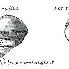
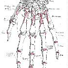
The scaphoid articulates with five bones: the radius, trapezoid, trapezium, lunate and capitate.
- proximal surface: radius
- distal surface: laterally with the trapezoid and trapezium; medially with the capitate
- ulnar surface: lunate
Proximally, the smooth convex surface of the scaphoid articulates with the distal radius.
The distal surface is split into two separate articular surfaces by a bony ridge. Radially, it articulates with the trapezoid and trapezium while on the ulnar side, it articulates with the capitate.
The medial surface has a concave appearance and articulates with the lunate.
Attachments
Musculotendinous
There are no musculotendinous attachments to the scaphoid bone.
Ligamentous
- dorsal surface: dorsal radiocarpal ligament
- radial surface: radial collateral ligament
- scapholunate ligament
- radioscapholunate ligament
- scaphocapitate ligament
- transverse carpal ligament
Relations
The radial artery crosses the dorsal surface of the scaphoid.
The scaphoid forms the radial portion of the carpal tunnel and is therefore related to the structures that pass through it, namely fibers from flexor digitorum profundus and superficialis, the median nerve, flexor pollicis longus and flexor carpi radialis.
Also located in the vicinity are the muscles of the thumb and associated tendons.
Arterial supply
Approximately 75% of the arterial supply is from branches of the radial artery through vascular perforations on the dorsal surface near the tubercle and waist . As the vascular supply to the proximal pole is mainly retrograde, a fracture through the tubercle or the waist places the proximal pole at risk of avascular necrosis.
Variant anatomy
- bipartite scaphoid
- tripartite scaphoid
- coalition with neighboring bones (rare)
- scaphoid hypoplasia (as long as not associated with other skeletal dysplasias)
Radiographic features
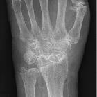
Plain radiograph
The scaphoid is the most radial of the proximal row of carpals, articulating with the distal radius, lunate and capitate. On the lateral view, it is projected through the carpus.
Development
Ossification
The scaphoid has a single ossification center, as do the trapezoid and trapezium. Ossification begins around the 4 year, and as with most ossification in the hand and wrist, it tends to occur earlier in females.
History and etymology
The term derives from the Ancient Greek word σκαφη (skaphe) meaning boat .
Related investigations
Plain radiograph
The scaphoid may be visualized on a number of series of the distal upper limb including:
Cross-sectional imaging
- CT scaphoid
- MRI scaphoid
Nuclear medicine
- scaphoid bone scan
Related pathology
- scaphoid fracture
- scaphoid non-union
- scapholunate advanced collapse
- scaphoid avascular necrosis
- idiopathic scaphoid avascular necrosis
- scapholunate instability
- scapholunate dissociation
- carpal tunnel syndrome
- scaphoid hypoplasia
- distal radial fracture
Siehe auch:
- Aseptische Knochennekrose
- Os trapezium
- Os scaphoideum bipartitum
- Scaphoidfraktur
- scaphoid non-union advanced collapse
- Fraktur
- Scaphoidpseudarthrose
- Tabatiere
und weiter:


 Assoziationen und Differentialdiagnosen zu Os scaphoideum: