shoulder osteoarthritis


Osteoarthritis of the shoulder or glenohumeral osteoarthritis is referred to progressive damage of the glenohumeral cartilage associated with bony erosions pain and a loss in function of the glenohumeral joint.
Epidemiology
The glenohumeral joint is one of the less common joints affected by osteoarthritis . Estimated radiographic prevalence is in the 16-20% range in an elderly population .
Risk factors
The main risk factor for glenohumeral osteoarthritis is age .
Other factors that increase the likelihood of developing osteoarthritis of the shoulder are :
- female sex
- obesity
- caucasian race
- prior trauma
- rotator cuff tears
- glenohumeral instability
- crystalline arthropathy
- sickle cell disease
Clinical presentation
Typical symptoms are slowly progressive posterior or deep localized shoulder pain associated with a limited range of motion, stiffness and worse at night. Other symptoms might include locking, grinding and joint instability .
Pathology
Generally, osteoarthritis is characterized by progressive joint alteration, due to a combination of mechanical, inflammatory and metabolical factors not only affect the hyaline cartilage but also the surrounding tissues, including the subchondral bone, the joint capsule and the synovium as well as the ligaments and in case of the shoulder the rotator cuff. These alterations are thought to arise from an imbalance between destruction and repair of the affected tissues . Like in other diarthrodial joints glenohumeral osteoarthritis is associated with thickening of the subchondral bone plate and marginal osteophyte formation, which usually can be seen at the posterior glenoid rim and the central part of the humeral head first. Soft tissue changes associated with the condition are capsular thickening and contraction potentially leading to an internal rotation deficit and further eccentric posterior glenoid erosion .
Etiology
Causes for the development of glenohumeral osteoarthritis include the following :
- idiopathic/unknown
- previous trauma
- glenohumeral dislocation
- proximal humeral fractures
- avascular necrosis of the shoulder
- inflammatory joint disease, e.g. septic arthritis
- hemochromatosis, hemophilia
- crystalline arthropathy
- iatrogenic, e.g. multiple intra-articular steroid injections
Classification
Similar to other joints glenohumeral osteoarthritis of the shoulder can be classified into primary and secondary, depending on whether it is due to a known predisposing factor or not.
An important classification scheme in the workup of glenohumeral osteoarthritis with regard to the management is the Walch classification of glenoid morphology which was later modified by Bercik et al.
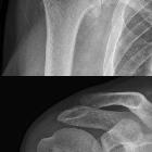
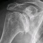
Radiographic features
General features are osteophyte formation, joint space narrowing and sclerosis of the subchondral bone plate. Subchondral cyst formation and remodeling of the articular surfaces or deformity are seen in more advanced stages.
Plain radiograph
Anteroposterior and lateral views of the shoulder are the main modality for the diagnosis and assessment.
Principal signs of osteoarthritis are the following:
- subchondral sclerosis
- joint space narrowing
- marginal osteophyte formation
- bony erosions/subchondral cyst formation
A radiological classification system generally used for the assessment of osteoarthritis is the Kellgren and Lawrence score which can also be used for the glenohumeral joint .
Specifically for the glenohumeral joint, a classification scheme originally suggested by Samilson and Prieto for glenohumeral osteoarthritis in the setting of glenohumeral instability has been used for grading due to its simplicity and reproducibility. It utilizes the size of inferior humeral osteophytes :
- grade 1: < 3mm
- grade 2: 3-7mm
- grade 3: >7mm
Ultrasound
Ultrasound can be used in the evaluation and workup of shoulder pain in particular shoulder impingement and rotator cuff tears, which represent an important differential diagnosis and critical criterium in surgical management.
CT
Computed tomography provides information on glenoid and proximal humeral anatomy and is invaluable for surgical planning in the assessment of the amount of bone stock.
MRI
In addition to the visualization of glenoid and humeral head morphology, MRI can help in the detection of underlying etiology. It allows the assessment of a variety of tissue abnormalities including cartilage, labrum and the glenohumeral ligaments. The capacity in the detection of cartilage injury is however limited if compared to other joints (e.g. knee hip or ankle) . In exchange, it provides valuable information in the evaluation of the rotator cuff, which forms an integral part in surgical planning.
Radiology report
The radiological report should include a description of the following:
- joint space narrowing and joint space width
- subchondral sclerosis
- presence and the location of osteophyte formation
- presence of subchondral cysts and/or bone erosion
- other findings e.g. subchondral fractures, signs of osteonecrosis
MRI
In addition to the above-mentioned features the MRI report should include the description of the following:
- cartilage and labral pathology
- subchondral bone marrow edema like signal
- joint effusion, synovitis
- intraarticular loose bodies
- rotator cuff pathology, in particular, massive and full-thickness rotator cuff tears and subscapularis tears
Treatment and prognosis
Management strategies include conservative non-surgical measures as well as surgical modalities and should be individualized with respect to the patient’s fitness and functional requirements .
Main objectivities are pain control and functional maintenance or restoration.
Non-operative management includes patient education, lifestyle and activity modifications and physical therapy as well as pain management with acetaminophen and nonsteroidal anti-inflammatory drugs.
Intra-articular injections of steroids and hyaluronic acid are other options .
Surgical management includes comprehensive arthroscopic management (cam procedure) and different options of shoulder arthroplasty from anatomic total shoulder arthroplasty over shoulder resurfacing arthroplasty or hemiarthroplasty to reverse total shoulder arthroplasty .
Indications for the different options depend on the age and activity of the patient, whether only the humeral head or both parts of the joints are involved and the integrity of the rotator cuff including the subscapularis tendon as well as humeral and glenoid morphology .
Differential diagnosis
The differential diagnosis of glenohumeral osteoarthritis includes the following :
- massive rotator cuff tear
- rotator cuff arthropathy
- acromioclavicular joint osteoarthritis
- glenohumeral instability
See also

