shoulder dislocation


The shoulder dislocation (more accurately termed a glenohumeral joint dislocation) involves separation of the humerus from the glenoid of the scapula at the glenohumeral joint.
This article contains a general discussion on shoulder dislocation. For specific dislocation types please refer to the following articles:
- anterior shoulder dislocation (95% of shoulder dislocations)
- posterior shoulder dislocation
- inferior shoulder dislocation
Epidemiology
Sex distribution is bimodal and relative incidence is dependent on patient age. Younger patients tend to be male and injury is often related to sporting trauma:
- younger: 20-30 years (male to female ratio of 9:1)
- older: 60-80 years (female to male ratio of 3:1)
Clinical presentation
Patients present with severe pain and restriction of movement of the shoulder. The majority of people who present with a shoulder dislocation do so after trauma, e.g. sporting trauma, assault, seizure, falls.
It is useful to determine whether the dislocation is acute, chronic or recurrent.
Pathology
Etiology
The shoulder is exceptionally maneuverable and sacrifices stability to enable an increase in function. Its shallow glenoid fossa, relatively weak glenohumeral ligaments, and redundant capsule render it particularly susceptible to dislocation. It is the most commonly dislocated large joint; indeed, the most commonly dislocated joint in the body . Approximately half of major joint dislocations seen in emergency departments are of the shoulder .
Shoulder dislocation almost exclusively occurs following trauma. The shoulder is in its weakest position when it is abducted and externally rotated. Sporting injuries and motor vehicle collisions are common causes.
Increased incidence in patients who have had a previous shoulder injury, and particularly in those who have dislocated previously.
The process of dislocation is massively disruptive to the labrum, joint capsule, supporting ligaments, and muscles. This is particularly true of anterior dislocations where there can be an injury to the anterior capsule, anterior labrum, or biceps tendon, or a combination thereof.
Relevant anatomy
The glenoid is a saucer-shaped extension of the scapula. Its shape means that it offers limited bony support to the joint. The glenoid is augmented by the cartilaginous labrum with additional support from the joint capsule, surrounding ligaments and the muscles of the rotator cuff. The labrum, capsule and ligaments tend to be stronger in younger patients.
Type of dislocation
Shoulder dislocations are usually divided according to the direction in which the humerus exits the joint:
- anterior >95%
- subcoracoid (majority)
- subglenoid (1/3)
- subclavicular (rare)
- posterior 2-4%
- inferior (luxatio erecta) <1%
Radiographic features
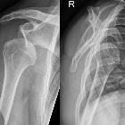
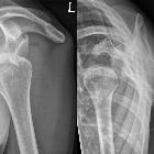
A shoulder x-ray series is sufficient in almost all cases to make the diagnosis, although CT and MR are often required to assess for the presence of subtle fractures of the glenoid rim or ligamentous/tendinous injuries respectively.
Plain radiograph
Anterior and inferior dislocations are usually simple diagnoses, with the humeral head and outline of the glenoid being incongruent.
Where the humeral head is displaced medially and overlies the glenoid, the dislocation is anterior.
Posterior dislocations can be difficult to identify on an AP view only (as may be obtained in the setting of a secondary survey of a trauma), as the humeral head moves directly posteriorly and congruency may appear to be maintained (at least at first glance).
All dislocations should be easily identified on trans-scapular Y views. When the humeral head is normally aligned, it will project centered over the center of the Y formed by the coracoid, blade of the scapula and spine of the scapula (acromion).
Report checklist
In addition to reporting the presence of a dislocation a number of features and associated findings should be sought and commented upon:
- direction of dislocation
- associated fractures/injuries
It is also important to remember to scrutinise the ribs and portion of the lungs and mediastinum included in the film for unexpected findings (e.g. pneumothorax). Think about the soft tissue structures that might be injured, particularly the neurovascular bundle with inferior dislocations.
Treatment and prognosis
The only treatment option for a dislocated shoulder is a prompt reduction. This is usually performed in the Emergency Department following sedation and appropriate analgesia. A number of techniques can be used to reduce the shoulder.
The ease of reduction is dependent on the age and build of the patient (younger, heavily built guys will be more difficult to reduce) and the time that the joint has been dislocated (the longer it has been out, the more difficult it is to get back in).
Rest is required following dislocation, so immobilization is required: three weeks for younger patients (<30 years old, who have a very high rate of recurrence) and 7-10 days in older patients. During this time gentle active motion should be carried out to preserve range of motion .
As a general rule, the shorter the duration of dislocation the fewer complications (size of Hill-Sachs lesion, neurovascular compromise, etc.).
Early arthroscopy, labral repair and debridement may be of use, especially in young patients with anterior dislocation in which there is a high (up to 85%) rate of recurrence .
Shoulder dislocations can also be associated with large rotator cuff tears in the older ages groups. The incidence starts to increase around age 40 and is especially high in patients above the age of 60 . The major morbidity associated with untreated massive rotator cuff tears in this age group requires a clinician to ensure actively that these injuries are not missed. This can be done by clinical examination, looking for weakness in the rotator cuff muscles or radiologically with ultrasound or MRI. Best outcomes are achieved with early surgical repair of the rotator cuff .
Differential diagnosis
- shoulder pseudodislocation: apparent inferior displacement of the humeral head from capsular distension secondary to a large hemarthrosis/effusion.
Siehe auch:
- hintere Schulterluxation
- Bankart-Läsion
- proximale Humerusfrakturen
- Hill-Sachs-Läsion
- Klavikulafraktur
- vordere Schulterluxation
- ossäre Bankart-Läsion
- luxatio erecta
- vordere Schulterinstabilität
- dislocated large joint
- chronische Schulterluxation
und weiter:


 Assoziationen und Differentialdiagnosen zu Schulterluxation:


