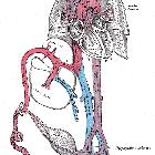
single umbilical artery


Single umbilical artery (SUA) results when there is a congenital absence of either the right or left umbilical artery. In the usual situation, there are paired umbilical arteries. For unknown reasons, the absence of the left umbilical artery is much more common (~70%).
Epidemiology
The estimated prevalence is ~0.4-1% of pregnancies . There may be an increased incidence of twin pregnancies and maternal diabetes.
Pathology
The occurrence of a single umbilical artery is thought to be due to secondary atresia or atrophy rather than primary agenesis of the artery. The remaining single artery is often quite large and approaches the size of the umbilical vein (which is usually larger than the artery).
In ~65% (range 57-75%) of cases, a single umbilical artery is present in isolation .
Associations
When found in isolation, it is usually not of clinical significance but there is an increased incidence of intrauterine growth restriction (IUGR) (~15%).
Recognized associations are thought to be present in ~35% (range 25-43%) of cases:
- lesser number of coils in the umbilical cord
- umbilical arterial aneurysm
- twin reversed arterial perfusion sequence
- increased incidence of intrauterine growth restriction (IUGR)
When found with other fetal anomalies, it can be also be associated with:
- chromosomal anomalies
- trisomy 21 (12.8% had an SUA), trisomy 18 (50% had an SUA), trisomy 13 (25% had an SUA)
- persistent right umbilical vein
- congenital renal anomalies
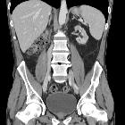
- renal agenesis: occurs usually on the side where the artery is absent
- sirenomelia
- velamentous insertion of the cord
Some suggest that complex congenital and chromosomal abnormalities are found almost exclusively when the left umbilical artery is absent
Radiographic features
Antenatal ultrasound
This is the imaging investigation of choice and an SUA is often detected incidentally on ultrasound. High-resolution ultrasound has a sensitivity and specificity approaching 100% . Sonographic features include:
- two vessels within the umbilical cord (one artery and one vein) instead of the usual three (best seen in cross-section)
- the single artery is often larger in caliber than normal and approaches the diameter of the accompanying vein
- examination of the fetal pelvis will demonstrate only one umbilical artery lateral to the bladder in its course toward the umbilical cord
Siehe auch:
- Pätau-Syndrom
- angeborene renale Anomalien
- Trisomie 18
- einseitige Nierenagenesie
- persistent right umbilical vein
- fetaler Blutkreislauf
- Insertio velamentosa
- Intrauterine Wachstumsretardierung
- chromosomale Anomalien
- Zwillingsschwangerschaft
- umbilical arterial aneurysm
- twin reversed arterial perfusion sequence
- lesser number of coils in umbilical cord
- Thrombose der Umbilikalarterien
- sirenomelia
- intra-uretine growth restriction (IUGR)
und weiter:


 Assoziationen und Differentialdiagnosen zu singuläre Nabelschnurarterie (sNSA):