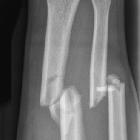
Ulna


The ulna (plural: ulnae) is one of the two long bones of the forearm, located medially in the supinated anatomic position. It has a larger proximal end and tapers to a smaller distal end (opposite to the radius).
Gross anatomy
Osteology
Prominent features of the ulna include:
- proximal: olecranon, trochlear notch, coronoid process, radial notch (lateral), sublime tubercle (medial)
- shaft: ulnar tuberosity
- distal: head, styloid process, fovea, groove for extensor carpi ulnaris
Articulations
- proximal (elbow joint)
- olecranon: ulnatrochlear joint (elbow flexion)
- radial notch: proximal radioulnar joint (site of supination and pronation)
- distal
- distal radioulnar joint
- wrist via the TFCC
Attachments
Musculotendinous
Anteriorly
- proximal:
- brachialis: ulnar tuberosity
- pronator teres (ulnar head): coronoid process
- flexor digitorum superficialis (humeroulnar head): coronoid process
- shaft:
- flexor digitorum profundus: proximal anterior surface ulna
- pronator quadratus: anterior surface of the distal quarter of the ulnar shaft
Posteriorly
- proximal:
- triceps brachii: proximal olecranon process
- anconeus: olecranon process
- shaft:
- supinator: posterior proximal shaft of ulna
- flexor carpi ulnaris (ulnar head): medial border olecranon + posterior border of ulna
- extensor carpi ulnaris: posterior border of ulna
- abductor pollicis longus: posterior surface of ulna
- extensor pollicis longus: middle third of the posterior surface of ulna
- extensor indicis: posterior surface of ulna
Ligamentous
- proximal:
- medial collateral ligaments of the elbow
- anterior band: inferior medial epicondyle to the sublime tubercle
- posterior band: medial epicondyle to the medial olecranon
- middle band (Transverse or Cooper's ligament): medial olecranon to the medial coronoid process
- anterior and posterior capsular ligaments of the elbow
- medial collateral ligaments of the elbow
- medial:
- anterior and posterior attachments of the annular ligament
- quadrate ligament
- oblique cord
- interosseous membrane
- distal:
- triangular fibrocartilage
- ulnar collateral ligament of the wrist
Relations and boundaries
The ulna and its attachments help to divide the forearm into anterior and posterior compartments.
Its subcutaneous border lies posteromedially and the antebrachial fascia attaches on either end.
Its interosseous border (anterolaterally) is attached to the interosseous membrane of the forearm.
Blood supply
The ulna is supplied by the ulnar artery and its continuation as the common interosseous artery with its branches, the anterior and posterior interosseous arteries.
Lymphatic drainage
Lymphatics of the hand and forearm drain either to the supratrochlear lymph node or directly into the lateral group of axillary lymph nodes.
Innervation
The periosteum is supplied anteriorly by the anterior interosseous nerve (branch of median nerve).
Posteriorly, the periosteum is supplied by the posterior interosseous nerve (branch of radial nerve).
Radiographic features
Carrying angle of the elbow of 15-20°. Increased in females.
Development
Intracartilaginous ossification begins in utero. Ossification centers include:
- shaft/diaphysis (8 weeks gestation)
- distal (5-7 years > 16-18 years)
- proximal (8-10 years > 13-15 years)
Ossification
Related pathology
- Monteggia fracture: fracture of ulna with dislocation of proximal radioulnar joint
- Galeazzi fracture: fracture of the radius with dislocation of distal radioulnar joint
- Essex-Lopresti fracture: fracture of the radial head with disruption of the interosseous membrane and dislocation of distal radioulnar joint
- Madelung deformity
- ulnotriquetrial abutment
Siehe auch:

 Assoziationen und Differentialdiagnosen zu Ulna:

