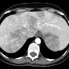
variköse Erweiterung der Pulmonalvenen

Imaging of
congenital lung diseases presenting in the adulthood: a pictorial review. Pulmonary varix. Curved multiplanar reconstruction (a) and 3D volume-rendered CT image (b) demonstrate pulmonary varix in a 73-year-old female

Pictorial
review of the pulmonary vasculature: from arteries to veins. Pulmonary vein varix. Axial contrast-enhanced CT image demonstrated a pulmonary vein varix (white arrow); note its proximity to the insertion in the left atrium. Varices can be saccular, tortuous, or confluent, with confluent being the most common
Pulmonary vein varix (PVV), also sometimes termed a pulmonary venous aneurysm, refers to a localized aneurysmal dilatation of a pulmonary vein. As it involves a venous structure, the former term is usually considered more appropriate. They are rare and may be congenital or acquired.
Clinical presentation
It is usually asymptomatic.
Pathology
While they can occur anywhere in the chest, they are thought to typically occur at the confluences of the veins adjacent to the left atrium .
They are sometimes classified into three morphological types:
- saccular type: considered most common
- tortuous type
- confluent type
Associations
Radiographic features
Plain radiograph
Non-specific and can present as a mass on a chest radiograph .
CTPA/contrast pulmonary angiography
Pulmonary angiography has been the mainstay of diagnosis . Allows accurate delineation of arterial and venous anatomy.
Siehe auch:

 Assoziationen und Differentialdiagnosen zu variköse Erweiterung der Pulmonalvenen:


