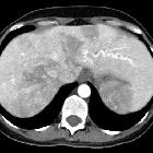
hereditary hemorrhagic telangiectasia


Hereditary hemorrhagic telangiectasia (HHT), also known as Osler-Weber-Rendu syndrome, is a rare inherited disorder characterized by abnormal blood vessel formation in the skin, mucous membranes, and organs including the lungs, liver, and central nervous system.
Epidemiology
Worldwide prevalence ~1.5 per 100,000. Wide geographic variability with a much higher incidence in certain regions, e.g. 1 in 200 in Dutch Antilles, 1 in 3500 in France.
Clinical presentation
Although the disease has a broad clinical spectrum, the classic clinical triad at presentation is epistaxis, multiple telangiectasias, and positive family history.
The diagnosis is a clinical diagnosis (Curacao criteria) based on the presence of 3 out of 4 of the following :
- recurrent spontaneous epistaxis
- multiple mucocutaneous telangiectasias
- characteristic sites include: oral cavity, lips, fingers and nose
- visceral AVMs
- first degree relative with HHT
Pathology
It is an autosomal dominant multi-organ vascular dysplasia, characterized by multiple arteriovenous malformations (AVMs) that lack an intervening capillary network. Telangiectasias (small superficial AVMs) are particularly common. Mutations have been found in one of several genes (three known so far). De novo mutations are rare, almost all have a first-degree relative affected.
Hereditary hemorrhagic telangiectasia can involve multiple organ systems. The spectrum includes:
- nasal: 90%
- telangiectasias of nasal mucosa
- complications: recurrent epistaxis
- skin and mucosal membranes: 90%
- telangiectasias of skin, oral cavity, conjunctivae
- complications: recurrent bleeding
- liver: 71-79%
- symptomatic liver involvement in HHT is uncommon but does occur; it has been attributed to three distinct clinical subtypes and is believed to be a consequence of the predominant hepatic shunt pattern
- high-output cardiac failure
- shunting that increases cardiac preload
- typically arteriovenous or portovenous shunts
- portal hypertension
- increased flow into the portal system (arterioportal shunt)
- hepatic anatomic abnormalities leading to increased intrahepatic resistance
- biliary disease
- shunting of the blood away from the peribiliary plexus (arteriovenous or arterioportal shunting)
- extensive arteriovenous shunting lead to biliary necrosis and bile leak
- complications: hepatomegaly, right upper quadrant pain, high-output cardiac failure, portal hypertension, mesenteric angina from steal phenomenon
- gastrointestinal tract: 20-40%
- AVMs or angiodysplasia in the stomach, small bowel or large bowel
- complications: recurrent GI bleeding
- pulmonary: 20%
- pulmonary arterio-venous malformations (AVMs)
- 36% of patients with solitary pulmonary AVM have HHT
- 57% of patients with multiple pulmonary AVMs have HHT
- complications
- pulmonary hemorrhage, hemoptysis (less common)
- complications of shunting (more common): paradoxical emboli (due to right to left shunt, e.g. stroke), septic emboli (e.g. cerebral abscess), hypoxemia, high-output cardiac failure
- pulmonary arterio-venous malformations (AVMs)
- CNS: 5-10%
- cerebral AVMs, spinal AVMs or cerebral aneurysms
- complications: headache, seizures, paraparesis, hemorrhage
- one-third of cerebral complications in HHT are due to cerebral AVMs or aneurysms, and two-thirds are due to paradoxical emboli from pulmonary AVMs
- increased incidence of capillary telangiectasia and developmental venous anomalies
Radiographic features
Imaging of visceral arteriovenous malformations
- lung
- chest x-ray: well-circumscribed mass (may be lobulated) with enlarged draining vein
- CT: well-circumscribed vascular mass with enhancing feeding artery and draining vein
- contrast echocardiography
- presence of contrast bubbles in the left atrium confirms the presence of a shunt
- characteristically, this occurs late (after several cardiac cycles), indicating a pulmonary shunt rather than intracardiac shunt
- CNS
- MR: cerebral and cerebellar AVMs typically in superficial locations
- gastrointestinal tract
- CT/CTA
- conventional angiography
- endoscopy
- capsule endoscopy
- nuclear medicine GI bleed study for active bleeding
- liver
- CT/CTA
- MRI
- conventional angiography
- ultrasound
Treatment and prognosis
Treatment of visceral lesions
- lung
- embolization; recanalization occurs in up to 20% post embolization
- surgical resection
- CNS
- embolization
- surgical resection
- stereotactic radiosurgery
- gastrointestinal tract
- embolization
- surgical resection
- endoscopic ablation/electrocautery
- liver
- embolization
- surgical resection
- liver transplantation
Prognosis
- most patients have a normal life expectancy
- 10% die of complications: usually stroke, cerebral abscess or massive hemorrhage
Siehe auch:
- zerebrale arteriovenöse Malformation
- arteriovenöse Malformationen der Lunge
- portale Hypertension
- spinale arteriovenöse Malformationen
- Morbus Osler ZNS-Manifestationen
- hereditäre hämorrhagische Teleangiektasie Leber
und weiter:
- kapilläre Teleangiektasien des ZNS
- Epistaxis
- variköse Erweiterung der Pulmonalvenen
- stroke in children and young adults
- bronchial arterial aneurysm
- Blue-rubber-bleb-naevus-Syndrom
- pathological conditions of hepatic vascularization
- Angiektasien
- Teleangiektasie
- pulmonary arteriovenous fistula in hereditary hemorrhagic telangiectasia
- brain abscess associated to Rendu-Osler-Weber disease
- pulmonary arteriovenous malformation in Rendu-Osler-Weber disease
- hereditary haemorrhagic telangiectasia: hepatic involvement
- Angiodysplasien des Gastrointestinaltraktes
- vaskuläre Syndrome
- arteriovenöse Fistel der Milz

 Assoziationen und Differentialdiagnosen zu Morbus Osler-Weber-Rendu:


