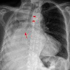
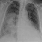
patterns of pulmonary opacification
Pulmonary opacification represents the result of a decrease in the ratio of gas to soft tissue (blood, lung parenchyma and stroma) in the lung. When reviewing an area of increased attenuation (opacification) on a chest radiograph or CT it is vital to determine where the opacification is. The patterns can broadly be divided into airspace opacification, lines and dots.
Classification of pulmonary opacification
- airspace opacification
- linear opacification
- reticular interstitial pattern, e.g. usual interstitial pneumonia
- reticulonodular interstitial pattern, e.g. sarcoidosis
- linear interstitial pattern, e.g. pulmonary edema
- nodular opacification
- miliary (<2 mm), e.g. miliary tuberculosis
- micronodular (2-7 mm), e.g. acute hypersensitivity pneumonitis
- nodule (7-30 mm), e.g. lung metastasis, lung granuloma
- mass (>30 mm), e.g. bronchogenic carcinoma
- pulmonary mucoid impaction, e.g. ABPA
See also
- parenchymal lung disease
- pulmonary opacification
- pulmonary lucency
Siehe auch:
- Atelektase
- Konsolidierung der Lunge
- Lungenkarzinom
- Sarkoidose
- Röntgen-Thorax
- Allergische bronchopulmonale Aspergillose
- Milchglasverschattungen
- Miliartuberkulose
- Lungenmetastasen
- gewöhnliche interstitielle Pneumonie (UIP)
- retikuläres Muster
- lung parenchyma
- parenchymal lung disease
- nodular opacification
- acute hypersensitivity pneumonitis
- Thorax Onlinekurs
- airspace nodules
- Bronchozele
- consolidation
- linear opacification
und weiter:

 Assoziationen und Differentialdiagnosen zu pulmonary opacity:
gewöhnliche
interstitielle Pneumonie (UIP)


