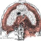
Lunge


The lungs are the functional units of respiration and are key to survival. They contain 1500 miles of airways, 300-500 million alveoli and have a combined surface area of 70 square meters (half a tennis court). Each lung weighs approximately 1.1 kg. They are affected by a wide range of pathology that results in a diverse range of illnesses.
Gross anatomy
The lungs lie in the thoracic cavity, separated by the heart and mediastinum. While the two lungs are similar, they are not completely symmetrical, having a different number of lobes and a different bronchial and vascular anatomy. In most individuals, the right lung is composed of three lobes subdivided into 10 segments and the left is composed of two lobes and eight segments. They are surrounded by the pleura which separates them from the chest wall.
See main article: bronchopulmonary segmental anatomy.
The lobes of the lungs are also incompletely separated by fissures. Each lung has an oblique fissure with a horizontal fissure also present in the right lung.
Tracheobronchial tree
The tracheobronchial tree is a branching structure of tubes of an ever-decreasing diameter that start at the larynx and end in the alveoli. It can broadly be divided into conduction and respiratory zones.
Conduction zone
Gas is warmed and humidified as it is conducted from the oropharynx to the functional portion of the lung where gas exchange occurs. The conduction zone is composed of the trachea, bronchi, bronchioles and terminal bronchioles. Their function is to optimize gas delivery to the functional portion of the lung. Their walls contain cilia to remove particulates from the inspired gas and cartilage to ensure that they do not collapse in expiration.
Respiratory zone
The respiratory zone is an extension of the tracheobronchial tree at the level of the terminal bronchioles. It is composed of respiratory bronchioles, alveolar ducts and alveoli, and is the location of gas transfer within the lung. In any organ, the parenchyma refers the functional tissues; thus, the strict definition of lung parenchyma only includes the alveoli, alveolar ducts, and respiratory bronchioles.
Blood supply
The lungs have dual arterial supply and venous drainage: pulmonary arteries and veins as well as bronchial arteries and veins:
Arterial supply
- pulmonary arteries: supply de-oxygenated blood from the right ventricle
- bronchial arteries: branches of the thoracic aorta that supply oxygenated blood
Venous drainage
- pulmonary veins: drain into the left atrium
- bronchial veins: drain to the pulmonary veins, superior vena cava, and azygos venous system
Innervation
The lungs are supplied by sympathetic, parasympathetic and visceral afferent fibers from the pulmonary plexus, which itself is composed of fibers from the vagus nerve (parasympathetic and visceral afferent fibers) and fibers from the upper four sympathetic ganglia.
- parasympathetic supply arises from the vagus nerve
- supplies afferent fibers for the cough reflex
- sensory fibers
- causes vasoconstriction of the pulmonary vasculature
- bronchoconstriction
- increases glandular secretion
- sympathetic supply arises from T2-6 sympathetic ganglia (via the pulmonary plexus)
- causes vasodilatation of the pulmonary vasculature
- bronchodilatation
- suppresses glandular secretion
Lymphatic drainage
The lymphatics drain via superficial and deep lymphatic plexuses to bronchopulmonary nodes at the hilum. The superficial lymphatic plexus is subpleural while the deep lymphatic plexus accompanies the bronchovascular structures with associated intrapulmonary lymph nodes.
Variant anatomy
- tracheal bronchus
- accessory fissures
- bronchopulmonary foregut malformations
- esophageal lung (extremely rare)
- main bronchus arises from the esophagus
- pulmonary vascular supply
- pulmonary aplasia
- horseshoe lung
Physiology
Lung function can be controlled externally via innervation of the respiratory muscles and diaphragm. However, control of the bronchi significantly affects respiratory function and gas exchange. For example,
- sympathetic innervation: norepinephrine acts on the beta receptors causing bronchodilation
- parasympathetic innervation: acetylcholine acts on M-1 muscarinic receptors and maintains the resting tone of the bronchiolar smooth muscle - this action is related to, but distinct from bronchoconstriction
- other non-autonomic nervous and biochemical stimuli including carbon dioxide and oxygen tension are also involved in the regulation process
Related pathology
- airway disease
- interstitial diseases
See also
Siehe auch:
- bronchopulmonary segments
- Pleura
- Zwerchfell
- left upper lobe apicoposterior segment
- parenchyma
- Mittellappen
- lung parenchyma
- linker Oberlappen
- Thorax Onlinekurs
- pulmonary opacity
- linker Unterlappen
- rechter Unterlappen
- rechter Oberlappen
- Lungenazinus
- right lung
- left lung
- apical segment
und weiter:

 Assoziationen und Differentialdiagnosen zu Lunge:


