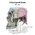
infratemporal fossa

The infratemporal fossa is a complex space of the face that lies posterolateral to the maxillary sinus and many important nerves and vessels traverse it. It lies below the skull base, between the pharyngeal sidewall and ramus of the mandible.
Gross anatomy
The infratemporal fossa is the space between the skull base, lateral pharyngeal wall, and the ramus of mandible. The fossa is actually open to the neck posteroinferiorly and in doing so has no true anatomical floor.
The fossa communicates with the temporal fossa via the space deep to the zygomatic arch, with the pterygopalatine fossa via the pterygomaxillary fissure, and with the middle cranial fossa via the foraminae ovale and spinosum.
The lateral pterygoid forms the foundation whereupon all other contents of the fossa are related. The branches of the mandibular nerve and the attachments of the medial pterygoid lie deep to the lateral pterygoid while the maxillary artery lies superficial to it. Between the two heads of the lateral pterygoid emerges the buccal branch of the mandibular nerve. The lingual and inferior alveolar branches of the mandibular nerve lie below the inferior border of the lateral pterygoid.
Boundaries
- medially: lateral pterygoid plate; tensor veli palatini and levator veli palatini muscles; superior constrictor muscle
- laterally: ramus and condylar process of mandible and zygomatic arch
- anteriorly: posterolateral wall of the maxillary sinus
- posteriorly: carotid sheath
- floor: medial pterygoid muscle
- roof: greater wing of sphenoid; foramen ovale; foramen spinosum
Relations
The infratemporal fossa encompasses the retroantral fat and medial parts of the following spaces :
Contents
- medial and lateral pterygoid muscles
- temporalis muscle
- maxillary artery and branches
- pterygoid venous plexus
- mandibular nerve and its branches (including lingual nerve)
- chorda tympani nerve
- posterior superior alveolar nerve of maxillary nerve
Siehe auch:

