pterygopalatine fossa

Pterygopalatine
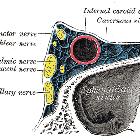
fossa • Pterygopalatine fossa (diagram) - Ganzer Fall bei Radiopaedia

Pterygopalatine
fossa • Pterygopalatine fossa: 3D diagram - Ganzer Fall bei Radiopaedia

Pterygopalatine
fossa • Pterygopalatine fossa - Ganzer Fall bei Radiopaedia

Pterygopalatine
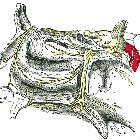
fossa • Pterygopalatine ganglion (illustration) - Ganzer Fall bei Radiopaedia

Pterygopalatine
fossa • Pterygopalatine fossa - 3D printed model - Ganzer Fall bei Radiopaedia

Pterygopalatine
fossa • Pterygopalatine fossa (annotated CT) - Ganzer Fall bei Radiopaedia
The pterygopalatine fossa (PPF), less commonly known as the sphenopalatine fossa, is a small but complex space of the deep face in the shape of an inverted pyramid located between the maxillary bone anteriorly, the pterygoid process posteriorly, and orbital apex superiorly. Its importance lies as the neurovascular crossroad of the nasal cavity, masticator space, orbit, oral cavity, and middle cranial fossa.
Gross anatomy
Borders
The walls of the pterygopalatine fossa are as follows:
- medial: perpendicular plate of the palatine bone
- lateral: narrowing to the pterygomaxillary fissure
- anterior: posterior wall of the maxillary sinus
- posterior: sphenoid bone (pterygoid process and inferior aspect of the greater wing)
- roof: incompletely formed by the greater wing of the sphenoid bone; inferior orbital fissure
- floor: narrowing to palatine canals; pyramidal process of the palatine bone
Communications
The pterygopalatine fossa is an important pathway for the spread of neoplastic and infectious processes:
- medially: communicates with the nasal cavity via the sphenopalatine foramen, which transmits the sphenopalatine artery, the nasopalatine nerve and the posterior superior nasal nerves
- laterally: communicates with the masticator space (or infratemporal fossa) via the pterygomaxillary fissure
- anteriorly: communicates with the orbit via the inferior orbital fissure (superiorly)
- posteriorly and superiorly: communicates with the Meckel cave and cavernous sinus (of the middle cranial fossa) via the foramen rotundum
- posteriorly and inferiorly: communicates with the middle cranial fossa via the vidian canal, which transmits the Vidian nerve
- posteriorly and medially: communicates with the nasopharynx via the palatovaginal canal, which transmits the pharyngeal nerve and the pharyngeal branch of maxillary artery
- inferiorly: communicates with the palate via the greater and lesser palatine canals (often these canals arise from the one canal, referred to as either the greater palatine canal or the pterygopalatine canal)
Contents
The pterygopalatine fossa contains fat and the following neurovascular structures:
- pterygopalatine ganglion
- maxillary artery (terminal portion), and its branches including the descending palatine artery
- emissary veins
- maxillary division of trigeminal nerve (Vb): enters via foramen rotundum
- nerve of the pterygoid canal
Radiological appearance
CT
- thin slice (<1 mm) bone algorithm reconstruction of non-contrast axial sections is the best approach to image the bony walls of the pterygopalatine fossa (see attached diagram)
MRI
- appears as a narrow cleft situated between the posterior wall of the maxillary sinus and the pterygoid plates
- T1W non-contrast axial images are the best approach for imaging the pterygopalatine fossa due to high fat-content
- contrast-enhanced images should also be acquired, as these are the best for evaluating the deep face
- mild post-contrast enhancement is normal due to the presence of small emissary veins
- may be obscured by susceptibility artifact in the following situations:
- "blooming" at the air-tissue interface between the maxillary sinus, which is a direct anterior relation
- dental fillings
Related pathology
Siehe auch:
und weiter:

 Assoziationen und Differentialdiagnosen zu Fossa pterygopalatina:


