Raumforderungen des Clivus

Radiological
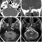
review of skull lesions. Chordoma. Axial head CT (a) shows an osteolytic destructive lesion involving the clivus (thick arrows) with extension into the posterior sphenoid sinus (arrowhead) and impression on the pons (curved arrow). Axial T1-weighted (b), axial T2-weighted (c) and sagittal post-contrast T1-weighted (d) images demonstrate an expansile mass centred at the clivus extending into the sphenoid sinus (arrowhead), left cavernous region (short, thick arrows) and partially encasing the left internal carotid artery. This mass enhances and displaces the pituitary gland (thin, long arrow) and has mass effect on the left aspect of the pons (curved arrow)

Radiological
review of skull lesions. Chondrosarcoma. Axial non-contrast CT (a) shows an osteolytic destructive lesion involving the right petro-occipital junction and clivus (arrowhead). Axial contrast-enhanced CT (b) demonstrates this lesion to have a soft-tissue component extending anteriorly into the sphenoid sinus (thick arrow) and posteriorly into the prepontine cistern with mass effect on the pons (dashed arrow). Axial (c) and coronal (d) CT angiogram images demonstrate involvement of the right petrous carotid canal (arrowheads) with the lesion surrounding the right internal carotid artery (thin arrow). Note the punctate calcifications (chondroid matrix) within the mass (curved arrow). Axial T1-weighted (e) and axial T2-weighted (f) images show a lobulated lesion involving the right petro-occipital junction and clivus (arrowhead). Note the extension of the lesion anteriorly into the sphenoid sinus (thick arrow) and posteriorly into the prepontine cistern with mass effect on the pons (dashed arrow). Axial SWAN (g) shows internal foci of low signal, consistent with calcifications (curved arrow). Post-contrast axial T1-weighted imaging (h) demonstrates heterogeneous (“whorls” of) enhancement (arrowhead)

Godtfredsen
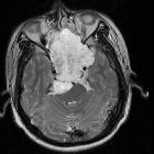
syndrome – recurrent clival chondrosarcoma with 6 years follow up: a case report and literature review. A Coronal view of the MRI brain shows residual tumour at the right basiocciput of the clivus. Enlarged photo shows tumour involving the right hypoglossal canal (in asterisk, *) Hypoglossal canal surrounded by jugular tubercle and occipital condyle resembles eagle head. (Eagle head image from Wikimedia Commons). B Axial view of MRI brain shows right clival mass at prepontine cistern causing compression the pons. C Residual clival chondrosarcoma extending to the right side sella compressing and displacing pituitary gland to the left

Clival
chordoma - CT and MRI. Coronal T2TSE. Heterogeneously hyperintense with cystic foci.

Clival
chordoma - CT and MRI. Sagittal T1+contrast. Heterogeneous “honeycombing” enhancement, with a predominantly peripheral distribution. Endocraneal invasion is evident, with pontine "thumbprinting".

Thumb sign
(chordoma) • Chordoma - clivus - Ganzer Fall bei Radiopaedia

Clival masses
• Pituitary adenoma - involving clivus - Ganzer Fall bei Radiopaedia

Clival masses
• Clival meningioma - Ganzer Fall bei Radiopaedia

Chondrosarcoma
• Chondrosarcoma - sphenoid wing - Ganzer Fall bei Radiopaedia

Clival masses
• Invasive nasopharyngeal carcinoma - with extension into the clivus - Ganzer Fall bei Radiopaedia
Raumforderungen des Clivus
Siehe auch:
- Tornwaldt-Zyste
- Chordom
- Tumoren der Hypophysenregion
- Makroadenom Hypophyse
- Multiples Myelom
- Ecchordosis physaliphora
- Kraniopharyngeom
- Mucocele
- Fibröse Dysplasie im Clivus
- Dural-Tail-Zeichen
- Nasopharynxkarzinom
- Chordom am Clivus
- normal bone marrow signal of the clivus
- Chondrosarkom der Schädelbasis
- Chondrosarkom des Clivus
- thumb sign of chordoma
- Meningeom des Klivus
- Osteomyelitis im Clivus
- Godtfredsen-Syndrom
- intrakranielle neuroenterische Zyste
und weiter:

 Assoziationen und Differentialdiagnosen zu Raumforderungen des Clivus:
intrakranielle
neuroenterische Zyste


