Parotistumoren


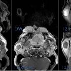
Warthin-Tumor
der Parotis, von links nach rechts: oben T2 FS ax, DWI ax, T2 FS cor; unten T1 ax, T1 FS KM ax und cor. Etwas Umgebungsreaktion um den Tumor ist nicht klassisch; typischerweise klar nach aussen abgrenzt.

Parotid gland
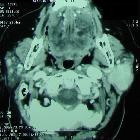
oncocytoma: a case report. Computer tomography (CT) findings: tumour mass of the left parotid gland.

Imaging of
parotid anomalies in infants and children. Acinic cell carcinoma. Satellite lymphadenomegaly on frontal fat sat T2-weighted image

Imaging of
parotid anomalies in infants and children. Intermediate-grade mucoepidermoid carcinoma: poorly delineated, heterogeneous tumor containing small necrotic areas [white arrow] and strongly enhanced, on ultrasonography (a and b), axial T2-weighted image (c) and T1-weighted image, after intravenous injection of gadolinium chelate (d). Time intensity curve with a signal growing rapidly in the first phase and a low wash out (< 30%): type C or plateau pattern (according to Yabuuchi), suggestive of malignancy (e). Conversely, time intensity curve of type B (orange line), with a high wash out (> 30%), in a case of parotid benign lymphadenopathy (f)

RETRACTED
ARTICLE: The diagnostic value of combined dynamic contrast-enhanced MRI (DCE-MRI) and diffusion-weighted imaging (DWI) in characterization of parotid gland tumors. Male patient, 62 years old presented with large Rt. Parotid swelling. a Axial T2 WI and b coronal T2 fat sat MRI showed large well-defined mass of high SI involving superficial and deep lobes of the Rt. parotid gland. It shows medial displacement of the Rt. parapharyngeal space. It is seen compressing and medially displacing the right CCA. The mass is seen contacting the right ramus of the mandible anteriorly and the right sternomastoid muscle posteriorly. c Coronal post contrast T1 fat sat MRI showed heterogeneous enhancement of the mass with cystic areas inside. d DWI the lesion appeared hypointense. e ADC map with mean ADC value = 2.2 × 10⁻³ mm2/s. f TIC showing type A curve (TTP = 140 s). Pathological diagnosis: pleomorphic adenoma

RETRACTED
ARTICLE: The diagnostic value of combined dynamic contrast-enhanced MRI (DCE-MRI) and diffusion-weighted imaging (DWI) in characterization of parotid gland tumors. Female patient 44 years old presented with right parotid swelling. a Axial T1-weighted MR image showed a well-defined lesion involving superficial lobe of right parotid gland. b Axial T2 and c coronal fat sat T2 WI showed heterogeneous SI of the right parotid lesion. d Post contrast T1 with fat suppression showed heterogeneous enhancement of the right parotid lesion. e DWI showed that lesion is hyperintense. f ADC map with mean ADC value = 0.78 × 10⁻³ mm2/s. g TIC showed type B curve (TTP = 80 S and WR > 30%). Pathological diagnosis: Warthin’s tumors

RETRACTED
ARTICLE: The diagnostic value of combined dynamic contrast-enhanced MRI (DCE-MRI) and diffusion-weighted imaging (DWI) in characterization of parotid gland tumors. Female patient, 40 years old presented with Rt. Parotid swelling. a Axial T1-weighted MRI showed large low SI infiltrating mass involving both superficial and deep lobes of the Rt. Parotid gland encasing the carotid sheath vessels. The mass extended to the Rt. parapharyngeal space and RT side of the oropharynx, RT masticator space and retromolar region. b Coronal T2 showed that the mass has heterogeneous high SI. c Axial post contrast T1 with fat suppression MRI: the lesion showed diffuse heterogeneous enhancement. d DWI showing the lesion was hyperintense. e ADC map with mean ADC value = 2 × 10⁻³ mm2/s. f TIC showing type C curve (TTP = 90 S, WR < 30%). Pathological diagnosis: acinic cell carcinoma.
Parotistumoren
Siehe auch:
- pleomorphes Adenom der Glandula parotis
- Hämangiom
- Pleomorphes Adenom
- Warthin-Tumor
- Tumoren der Speicheldrüsen
- Lymphangiom
- Adenoid-zystisches Karzinom
- zystische Läsionen der Glandula parotis
- myoepithelioma
- minor salivary gland tumour
- Vergrößerung der Glandula parotis
- Lipom der Speicheldrüsen
- Hämangiom Glandula parotis
- Lipom der Glandula parotis
- Speicheldrüsenmetastasen
- acinic cell carcinoma of salivary glands
- Teratom der Glandula parotis
- parotid adenocarcinoma
- staging of malignant salivary gland tumours
- Biopsie Glandula parotis
- Lungenkarzinome vom Speicheldrüsentyp
- mukoepidermoides Karzinom
- parotid lymphangioma
- intraparotid neurofibroma
- mukoepidermoides Karzinom der Parotis
- maligne Tumoren der Parotis
- infantiles Hämangiom der Glandula parotis
- intraparotideale Lymphknoten
- acinic cell carcinoma of the parotid gland
- enhancing mass in parotid gland
- intraparotideale Metastasen
- Abszess der Glandula parotis
- Adenoid-zystisches Karzinom der Speicheldrüsen
- infantile Parotistumoren
- parotid sarcoidosis
und weiter:

 Assoziationen und Differentialdiagnosen zu Parotistumoren:
Lungenkarzinome
vom Speicheldrüsentyp
Adenoid-zystisches
Karzinom der Speicheldrüsen


