spinal cord compression

Epidurale
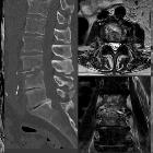
Lipomatose der BWS mit hochgradiger Einengung des Duralsackes bei gleichzeitig Vorwölbung der Wirbelkörperhinterkante durch metastatische Durchsetzung mit pathologischer Fraktur. Links Computertomografie, rechts folgend MRT sagittal T2 und T1 sowie 3 Schichten axial T2. In der Computertomografie schon geringe Höhenminderung von BWK 5 und nach dorsal vorwölbender Weichteilanteil der Metastase in BWK 6, in der MRT ca. 4 Wochen später zunehmender Einbruch BWK 5 mit deutlicher Pelottierung des Myelons. Die durale Enge wird durch das dorsale langstreckig epidurale liegende Fett trotz des knöchern eher weiten Spinalkanals verschärft. Weitere Metastasen in zahlreichen anderen Höhen.

Spinal cord
compression • Spinal cord compression from cervical disc bulge - Ganzer Fall bei Radiopaedia

Spinal cord
compression • Spondylolisthesis - Ganzer Fall bei Radiopaedia

Spinal
epidural abscess • Epidural abscess of the cervical spine - Ganzer Fall bei Radiopaedia

Spinal cord
compression • Discitis osteomyelitis - Ganzer Fall bei Radiopaedia

Neurofibromatosis
type 1 • Neurofibromatosis type 1 - Ganzer Fall bei Radiopaedia

Spinal cord
compression • Dens fracture and epidural hematoma - Ganzer Fall bei Radiopaedia

Spinal cord
compression • Cervical canal stenosis with cord compression - Ganzer Fall bei Radiopaedia

Spinal cord
compression • Metastatic spinal cord compression - Ganzer Fall bei Radiopaedia

Spinal cord
compression • Metastatic renal cell carcinoma with cord compression - Ganzer Fall bei Radiopaedia

Spinal cord
compression • Cord compression due to osseous metastases - Ganzer Fall bei Radiopaedia

Spinal cord
compression • Metastatic cord compression - Ganzer Fall bei Radiopaedia

Spinal cord
compression • Metastatic cord compression - Ganzer Fall bei Radiopaedia

Spinal cord
compression • Metastatic thoracic cord compression: prostate carcinoma - Ganzer Fall bei Radiopaedia

Minimally
displaced unilateral facet fracture of cervical spine can lead to spinal cord injury: a report of two cases. Magnetic resonance imaging using a sagittal short T1 inversion recovery (STIR) sequence and b axial T2 weighted image at C5/6 at re-admission shows disc injury and spinal cord compression by a posterior epidural mass accompanied by an intramedullary signal intensity change at this level. Prevertebral soft-tissue edema, injury of the interspinous ligament, and a narrowed canal also are evident. There is no flow void in right vertebral artery on axial T2 weighted image, suggesting vertebral artery injury
Spinal cord compression is a surgical emergency, usually requiring prompt surgical decompression to prevent permanent neurological impairment. If the spinal roots below the conus medullaris are involved, it is termed cauda equina syndrome.
Pathology
Etiology
There are numerous causes of cord compression. These can be divided according to the location of the compressing mass:
- intervertebral disc
- disc protrusion/disc extrusion
- diskitis osteomyelitis (usually associated with an epidural abscess)
- degenerative anterolisthesis/spondylosis
- vertebral
- trauma
- vertebral fracture (e.g. burst fracture)
- fracture-dislocation
- tumor
- vertebral metastasis
- primary vertebral tumor (e.g. aggressive vertebral hemangioma)
- trauma
- epidural space
- dura
- intradural space
- nerve sheath tumor (spinal schwannoma or neurofibroma)
Lesions of the cord itself can present in a similar manner to extrinsic cord compression but are usually considered separately (e.g. spinal cord tumors, spinal cord abscess).
Siehe auch:
- vertebrale Metastasen
- Spinalkanalstenose
- Bandscheibenprotrusion
- spinale Schwannome
- spinale Arachnoidalzyste
- Spondylodiscitis
- Neurofibrom
- Bandscheibenextrusion
- Berstungsfraktur
- intraspinales Meningeom
- Myelopathie durch spinale Enge
- zervikale Myelopathie
- Myelonkompression durch Metastasen
und weiter:

 Assoziationen und Differentialdiagnosen zu Myelonkompression:


