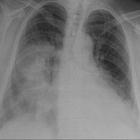
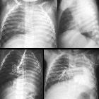
hemithorax white-out (differential)
Complete white-out of a hemithorax on the chest x-ray has a limited number of causes. The differential diagnosis can be shortened further with one simple observation: the position of the trachea. Is it central, pulled or pushed from the side of opacification? Is there pulmonary volume loss or volume gain? Is there rib crowding? Is the heart pushed or pulled?
Trachea pulled toward the opacified side
- pneumonectomy
- total lung collapse: e.g. endobronchial intubation
- pulmonary agenesis
- pulmonary hypoplasia
Trachea remains central in position
- consolidation
- pulmonary edema/ARDS
- pleural mass: e.g. mesothelioma
- chest wall mass: e.g. Askin/Ewing sarcoma
- being on extracorporeal membrane oxygenation (ECMO)
Pushed away from the opacified side
See also
Siehe auch:
- Konsolidierung der Lunge
- Pleuraerguss
- Lungenödem
- acute respiratory distress syndrome (ARDS)
- Zwerchfellhernie
- Mesotheliom
- Lungenhypoplasie
- konsolidierte Pneumonie
- Tubus im rechten Hauptbronchus
- pulmonale Aplasie
- pulmonale Raumforderung
- total lung collapse
- Askin / Ewing sarcoma
und weiter:

 Assoziationen und Differentialdiagnosen zu komplette Verschattung Hemithorax:


