ophthalmoplegia
Ophthalmoplegia describes the abnormal eye movement that occurs because of paralysis of one or more of the six extraocular muscles involved in eye movements. Classification can be based on the cause of the ophthalmoplegia or the directions of the affected movements.
There are numerous causes of ophthalmoplegia that relate to deficiency or disease in any part of the system:
- globe
- muscle dysfunction: progressive external ophthalmoplegia, Kearns-Sayre syndrome, Graves disease
- neuromuscular junction: myasthenia gravis
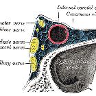
- cranial nerves: cavernous sinus syndrome, raised intracranial pressure (ICP)
- brainstem nuclei: brainstem stroke e.g. Foville syndrome
- white matter tracts: internuclear ophthalmoplegia
- dorsal midbrain: Parinaud syndrome
- cerebral cortex: stroke
- toxin: venom
- deficiency: thiamine deficiency in Wernicke encephalopathy
The cranial nerves involved in eye movement by innervation of the extraocular muscles are:
- oculomotor nerve (CN III): innervates medial rectus muscle, superior rectus muscle, inferior rectus muscle, inferior oblique muscle, pupilloconstrictor muscle and the striated portion of the levator palpebrae superioris muscle
- trochlear nerve (CN IV): innervates superior oblique muscle
- abducens nerve (CN VI): innervates lateral rectus muscle
Siehe auch:
- Korsakow-Syndrom
- Morbus Basedow
- Musculus obliquus inferior
- Nervus abducens
- Apoplex
- Nervus oculomotorius
- Musculus obliquus superior
- Kearns-Sayre-Syndrom
- Aquäduktsyndrom
- Nervus trochlearis
- Musculus rectus lateralis
- äußere Augenmuskulatur
- Internukleäre Ophthalmoplegie
- Musculus rectus medialis
- Musculus rectus superior
- Musculus rectus inferior
- Augenbewegungen

 Assoziationen und Differentialdiagnosen zu Ophthalmoplegie:


