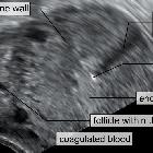
Ectopic


Ectopic pregnancy refers to the implantation of a fertilised ovum outside of the uterine cavity.
Epidemiology
The overall incidence has increased over the last few decades and is currently thought to affect 1-2% of pregnancies. The risk is as high as 18% for first trimester pregnancies with bleeding . There is an increased incidence associated with in-vitro fertilisation pregnancies.
Clinical presentation
The classic presentation is with abdominal pain and bleeding. In practice, the symptoms are not necessarily severe - often there may be only mild pelvic pain and spotting in early pregnancy (5-9 weeks of amenorrhea ). Nonetheless, monitoring of hemodynamic status is crucial, as hemorrhage can be life-threatening.
Pathology
Locations
In the vast majority of cases, the ectopic implantation site is within a Fallopian tube.
- tubal ectopic: 93-97%
- ampullary ectopic: most common ~70% of tubal ectopics and ~65% of all ectopics
- isthmal ectopic: ~12% of tubal ectopics and ~11% of all ectopics
- fimbrial ectopic: ~11% of tubal ectopics and ~10% of all ectopics
- interstitial ectopic/cornual ectopic: 3-4%; also essentially a type of tubal ectopic
- ovarian ectopic: ovarian pregnancy; 0.5-1%
- cervical ectopic: cervical pregnancy; rare <1%
- scar ectopic: site of previous Cesarian section scar; rare
- abdominal ectopic: rare ~1.4%
Risk factors
- in vitro fertilisation
- prior ectopic pregnancy
- tubal injury or surgery, including tubal ligation
- pelvic inflammatory disease
- salpingitis isthmica nodosa
- endometrial injury
- intrauterine contraceptive devices
- endometriosis
- previous placenta previa
- congenital uterine anomalies
- smoking
- past history of spontaneous or induced abortions
- maternal age (advanced maternal age increases the risk of ectopic pregnancy)
- history of subfertility
Markers
Serum beta HCG levels tend to increase at a slower rate. Whereas a normal doubling rate in early pregnancy is approximately 48 hours, an increase of 50% or less in 48 hours is strongly suggestive of a non-viable (either intra- or extrauterine) pregnancy . Rarely the urinary and/or serum b-HCG will be negative despite an ectopic pregnancy .
Serum progesterone levels are generally lower in a non-viable (including ectopic) pregnancy ; progesterone of 5 ng/ml or less is strongly associated with pregnancy failure, whereas in a viable pregnancy, progesterone is usually 20 ng/ml or more . Clearly, there is a significant grey zone. Furthermore, serum progesterone levels may take days to process. Progesterone is therefore not included in standard protocols for managing the suspected ectopic pregnancy.
Radiographic features
It is useful to know a quantitative beta HCG prior to scanning as this will guide what you expect to see. At levels <2000 IU, a normal early pregnancy may not be visible.
The most reliable sign of ectopic pregnancy is the visualization of an extra-uterine gestation, but this is not seen in 15-35% of ectopic pregnancies .
Ultrasound
The ultrasound exam should be performed both transabdominal and transvaginally. The transabdominal component provides a wider overview of the abdomen, whereas a transvaginal scan is important for diagnostic sensitivity.
Positive sonographic findings include:
- uterus
- an empty uterine cavity or no evidence of an intrauterine pregnancy
- an exception to this is a rare heterotopic pregnancy
- pseudogestational sac or decidual cyst: may be seen in 10-20% of ectopic pregnancies
- current evidence suggests that one should not initiate treatment for ectopic pregnancy in a haemodynamically stable woman on the basis of a single hCG value
- decidual cast
- thick echogenic endometrium
- an empty uterine cavity or no evidence of an intrauterine pregnancy
- tube and ovary
- simple adnexal cyst: 10% chance of an ectopic
- complex extra-adnexal cyst/mass: 95% chance of a tubal ectopic (if no IUP)
- an intra-adnexal cyst/mass is more likely to be a corpus luteum
- solid hyperechoic mass is possible but non-specific
- tubal ring sign
- 95% chance of a tubal ectopic if seen
- described in 49% of ectopics and in 68% of unruptured ectopics
- ring of fire sign: can be seen on color Doppler in a tubal ectopic, but can also be seen in a corpus luteum
- an absence of color Doppler flow does not exclude an ectopic
- live extrauterine pregnancy (i.e. extra-uterine fetal cardiac activity): 100% specific, but only seen in a minority of cases
- peritoneal cavity
- free pelvic fluid or hemoperitoneum in the pouch of Douglas
- the presence of free intraperitoneal fluid in the context of a positive beta HCG and the empty uterus is
- ~70% specific for an ectopic pregnancy
- ~63% sensitive for ectopic pregnancy
- not specific for ruptured ectopic (seen in 37% of intact tubal ectopics)
- the presence of free intraperitoneal fluid in the context of a positive beta HCG and the empty uterus is
- free fluid in the hepatorenal recess
- interrogation of the right upper quadrant for free fluid reduces time to diagnosis
- free fluid in Morison's pouch in the context of an ectopic pregnancy is highly suggestive that operative management will be necessary
- live pregnancy: 100% specific, but only seen in a minority of cases
- free pelvic fluid or hemoperitoneum in the pouch of Douglas
In patients receiving in vitro fertilisation (IVF), it is important not to be completely reassured by the presence of a live intrauterine pregnancy , as there is a possibility of a coexisting ectopic pregnancy in ~1-3:100 (i.e. heterotopic pregnancy). In patients not receiving IVF, the risk of heterotopic pregnancy is minuscule (1:30,000).
Complications
Complications somewhat depend on the type of ectopic. General complications for a typical (tubal) ectopic pregnancy include:
- tubal rupture: 15-20%
Treatment and prognosis
Management depends on the location of the ectopic pregnancy and the patient's hemodynamic status. In general, the options are:
- surgical: (in the case of tubal ectopics with open or laparoscopic salpingectomy or salpingotomy)
- medical
- methotrexate (a folate antagonist) either administered systemically or by direct ultrasound-guided injection
- relative contraindications to methotrexate include :
- rupture
- mass >3.5 cm
- fetal cardiac activity
- bHCG >6000-15,000 mIU/mL
- the gestational mass can paradoxically increase in size following methotrexate on subsequent scanning and does not necessarily imply failure of methotrexate therapy
- potassium chloride (via ultrasound-guided direct injection only)
- conservative or expectant management is being recognized as an option for those ectopics where rupture has not occurred (i.e. no hemoperitoneum) and fetal demise has already taken place
Differential diagnosis
The differential diagnosis of abdominal pain in a pregnant patient is broad. An ectopic pregnancy must be excluded by ultrasound. Other common diagnoses in this setting include:
- ruptured corpus luteum
- exophytic corpus luteum of pregnancy
- intrauterine pregnancy
- incidental adnexal mass
- appendicitis (negative beta-hCG)
The scenario of clinically suspected ectopic pregnancy that is not confirmed on ultrasound, is referred to as a pregnancy of unknown location, with the alternative possibilities being of very early pregnancy or a completed miscarriage.
Siehe auch:
- tubal ectopic pregnancy
- interstitial ectopic pregnancy
- live ectopic pregnancy
- pregnancy of unknown location
- ampullary ectopic pregnancy
- isthmic ectopic pregnancy
- Eileiterschwangerschaft
- akuter Beckenschmerz in der Schwangerschaft
- blob sign of ectopic pregnancy
- Extrauteringravidität in Kaiserschnittnarbe
- gleichzeitige intrauterine und ektope Schwangerschaft
- Extrauteringravidität in der Leber
und weiter:
- double decidual sac sign
- adnexal ectopic pregnancy
- per vaginal bleeding in the exam
- Akutes Abdomen
- first trimester
- cornual ectopic pregnancy
- cervical ectopic pregnancy
- ovarian ectopic pregnancy
- differential diagnosis for bleeding in pregnancy
- decidual reaction
- Schwangerschaft im Tubenwinkel
- collaborative publications by radiopaedians
- Corpus luteum Zyste
- empty gestational sac
- decidual cyst
- decidual cast
- tubal ring sign
- angular ectopic pregnancy
- Lithopädion
- akuter Beckenschmerz
- abdominelle Extrauteringravidität
- Schwangerschaft mit liegender Spirale
- atypische Extrauteringravidität

 Assoziationen und Differentialdiagnosen zu Extrauteringravidität:


